#orthopedi #conference#Arteries Of The Hand#Deep Palmar Arch #Superficial Palmar Arch#Common Digital Arteries #Digital Arteries to the Thumb#Proper Digital Arteries to the Fingers#neck#pain#orthopedic #ortho#orthopedic#osteoarthritis#hip#Pain #Stiffness#Tenderness#Loss of flexibility Grating sensation#Bone spurs #Swelling#common #conference.#Orthopedics#Orthopedic surgery#Joints#Bone#Arthritis #fracture #Dislocation#Sprain#Strain#$Tendinitis#Bursitis#Osteoporosis #scoliosis #kyphosis #lordosis #Spine#Back pain#Neck pain #hip #Knee#Shoulder#Elbow#Wrist#Hand#Foot#Ankle#Prosthetics #orthotics s#Physical therapy#Rehabilitation #Sports medicine#Trauma#Joint replacement#Total joint reconstruction #Arthroscopy#Soft tissue repair#Cartilage restoration#Joint preservation #Orthopedic implants#biomaterials #Orthopedic devices#Orthopedic equipment#Orthopedic instruments#Orthopedic braces.#Orthopedics #Orthopedic surgery#Joint replacement#Arthroplasty#Hip replacement #Knee replacement#Shoulder replacement#Spine surgery #Scoliosis#Deformity correction#Fracture
Saturday, April 29, 2023
4th Edition of Orthopedics summit and Sports Medicine 20-22 June 2023 | ...
#orthopedi #conference#Arteries Of The Hand#Deep Palmar Arch #Superficial Palmar Arch#Common Digital Arteries #Digital Arteries to the Thumb#Proper Digital Arteries to the Fingers#neck#pain#orthopedic #ortho#orthopedic#osteoarthritis#hip#Pain #Stiffness#Tenderness#Loss of flexibility Grating sensation#Bone spurs #Swelling#common #conference.#Orthopedics#Orthopedic surgery#Joints#Bone#Arthritis #fracture #Dislocation#Sprain#Strain#$Tendinitis#Bursitis#Osteoporosis #scoliosis #kyphosis #lordosis #Spine#Back pain#Neck pain #hip #Knee#Shoulder#Elbow#Wrist#Hand#Foot#Ankle#Prosthetics #orthotics s#Physical therapy#Rehabilitation #Sports medicine#Trauma#Joint replacement#Total joint reconstruction #Arthroscopy#Soft tissue repair#Cartilage restoration#Joint preservation #Orthopedic implants#biomaterials #Orthopedic devices#Orthopedic equipment#Orthopedic instruments#Orthopedic braces.#Orthopedics #Orthopedic surgery#Joint replacement#Arthroplasty#Hip replacement #Knee replacement#Shoulder replacement#Spine surgery #Scoliosis#Deformity correction#Fracture
Friday, April 28, 2023
What is Anterior Longitudinal Ligament?
As an AI language model, I do not have the ability to browse social media platforms like Instagram and view the content posted there. However, I can provide you with some general information about the Anterior Longitudinal Ligament (ALL) if that's what you're asking.
The ALL is a long, strong band of connective tissue that runs vertically along the front surface of the vertebral bodies in the spine. It helps to support and stabilize the spine, as well as limit excessive movement of the vertebral column. Injuries to the ALL can occur from trauma, such as a car accident or a fall, and can result in spinal instability and other complications.
Thursday, April 27, 2023
What is chemotherapy and types.
chemotherapy##Arteries Of The Hand#Deep Palmar Arch #Superficial Palmar Arch#Common Digital Arteries #Digital Arteries to the Thumb#Proper Digital Arteries to the Fingers#neck#pain#orthopedic #ortho#orthopedic#osteoarthritis#hip#Pain #Stiffness#Tenderness#Loss of flexibility Grating sensation#Bone spurs #Swelling#common #conference.#Orthopedics#Orthopedic surgery#Joints#Bone#Arthritis #fracture #Dislocation#Sprain#Strain#$Tendinitis#Bursitis#Osteoporosis #scoliosis #kyphosis #lordosis #Spine#Back pain#Neck pain #hip #Knee#Shoulder#Elbow#Wrist#Hand#Foot#Ankle#Prosthetics #orthotics s#Physical therapy#Rehabilitation #Sports medicine#Trauma#Joint replacement#Total joint reconstruction #Arthroscopy#Soft tissue repair#Cartilage restoration#Joint preservation #Orthopedic implants#biomaterials #Orthopedic devices#Orthopedic equipment#Orthopedic instruments#Orthopedic braces.#Orthopedics #Orthopedic surgery#Joint replacement#Arthroplasty#Hip replacement #Knee replacement#Shoulder replacement#Spine surgery #Scoliosis#Deformity correction#Fracture
Monday, April 24, 2023
4th Edition of Orthopedic 20-22 June 2023 San Francisco,United States.
Award Nomination - https://x-i.me/ortawr21
Member Nomination - https://x-i.me/ortmem0
Abstract Sumission - https://x-i.me/ortaji1
#Arteries Of The Hand#Deep Palmar Arch #Superficial Palmar Arch#Common Digital Arteries #Digital Arteries to the Thumb#Proper Digital Arteries to the Fingers#neck#pain#orthopedic #ortho#orthopedic#osteoarthritis#hip#Pain #Stiffness#Tenderness#Loss of flexibility Grating sensation#Bone spurs #Swelling#common #conference.#Orthopedics#Orthopedic surgery#Joints#Bone#Arthritis #fracture #Dislocation#Sprain#Strain#$Tendinitis#Bursitis#Osteoporosis #scoliosis #kyphosis #lordosis #Spine#Back pain#Neck pain #hip #Knee#Shoulder#Elbow#Wrist#Hand#Foot#Ankle#Prosthetics #orthotics s#Physical therapy#Rehabilitation #Sports medicine#Trauma#Joint replacement#Total joint reconstruction #Arthroscopy#Soft tissue repair#Cartilage restoration#Joint preservation #Orthopedic implants#biomaterials #Orthopedic devices#Orthopedic equipment#Orthopedic instruments#Orthopedic braces.#Orthopedics #Orthopedic surgery#Joint replacement#Arthroplasty#Hip replacement #Knee replacement#Shoulder replacement#Spine surgery #Scoliosis#Deformity correction#Fracture
Thursday, April 20, 2023
Novel stem cell therapy for repair of knee cartilage
Mayo Clinic offers a unique regenerative medicine approach for repairing knee cartilage, which can be completed in a single surgery. The Food and Drug Administration approved the use of this technique, known as recycled cartilage auto/allo implantation (RECLAIM), in a trial utilizing the stem cell bank in the Mayo Clinic Center for Regenerative Biotherapeutics.
"Mayo is unique in having an adipose-derived allogeneic stem cell bank. It provides us with donor mesenchymal stem cells, which we mix with recycled autologous cells to quickly obtain enough cells to fill the patient's cartilage defect without operating twice," says Daniel B. Saris, M.D., Ph.D., an orthopedic surgeon at Mayo Clinic in Rochester, Minnesota, who specializes in knee surgery and focuses on regenerative medicine.
Dr. Saris previously performed the RECLAIM cartilage repair technique in Europe. "The results, about four years out, are very good — comparable to or better than other cell therapies, except these patients achieve normal function after surgery about six months more quickly," he says.
Planning is underway for a clinical trial at Mayo Clinic. RECLAIM is used to repair symptomatic cartilage defects, usually resulting from trauma or an athletic injury. The procedure might be suitable for nonarthritic patients ages 18 to 50 who have fresh cartilage defects.
Recycling patient cells
Existing cell therapy to repair knee cartilage generally involves surgically debriding the cartilage defect and then taking a biopsy of healthy cartilage from the patient. The biopsy is cultured in an outside laboratory, and the cultured cells are implanted weeks later. "We wanted to improve this technique because during the waiting period, the patient's life is on hold, costs increase and the logistics can be complex.
RECLAIM's innovation starts with saving the patient's debrided tissue. "That tissue is always a bit frilly and is normally discarded. "But we found that the cells in that tissue are still very viable. We recycle them."
The resected tissue is processed and, using a rapid isolation protocol, digested into chondrons. Mixing the chondrons with allogeneic stem cells from the stem cell bank provides sufficient cells to immediately re-inject into the patient.

"This is a highly innovative procedure.You have to find an intricate balance — loading enough cells to grow into healthy tissue but not overloading the space so the cells are squished when the patient starts rehab."
Most patients return home on the day of surgery. They generally need to wait nine to 12 months before a full return to sports; that interval provides time for the cartilage to grow and the patient to regain muscle control. "But apart from sports, patients can go back to normal life within days and physical activities within three to four months of surgery.
Mayo Clinic's multidisciplinary approach provides the range of care needed by patients at all stages of knee cartilage repair. Before surgery, advanced imaging helps pinpoint the cartilage defect. "Our physiotherapists and athletic trainers also determine prior to surgery how we can optimize the patient's musculoskeletal control and function, and then work with the patient on rehab after surgery," Dr. Saris says.
Mayo Clinic also has the breadth of orthopedic expertise to manage problems that patients often experience alongside damaged knee cartilage, such as varus deformity and anterior cruciate ligament or meniscus lesions. "If a cartilage repair procedure fails, it's generally because not enough attention was paid to other factors — the meniscus or the knee's alignment or stability. "Our unique multidisciplinary team looks at all aspects of a patient's care. Our chances of success for these complex biological reconstructions is therefore high."
The cartilage repair technique illustrates Mayo Clinic's commitment to applying regenerative medicine to orthopedic surgery.
More information: https://orthopedic-conferences.pencis.com/
#hip replacement#Vertebral body tethering#athletes#Soft Tissue#Common Types of Bone # Fracture s#stem cells#Back Surgery#conference #spine diseases #sports #injuries#degenerative diseases#congenital disorders#Lancet #Arthroplasty#Injury#Shoulder surgery#Elbow Surgery#Spine #bone #Knee#Physiotherapy#Foot Surgery#Ankle SurgeryA#Posture#orthopedicdoctor #Gait#Cartilage#Osteoarthritis #long bones# orthopedic #medicine #sports
Identifying best candidates with avascular necrosis for regenerative alternative to hip replacement

Avascular necrosis is also a rare side effect of heavy steroid use as part of some chemotherapy treatment. Left untreated, tiny fractures from avascular necrosis worsen, and sometimes the joint collapses. Without hip decompression surgery, 90% of people with avascular necrosis need a total hip replacement.
A great mystery of orthopedics
Avascular necrosis of the hip has been called one of the great mysteries of orthopedics. Once bone cells begin to die, nothing can stop the damage, unless it is caught early or there is intervention to try to restore the blood flow.
Mayo Clinic has been offering hip decompression surgery for over 15 years. This regenerative procedure taps the body's ability to heal decaying bone cells. A small hole is drilled in the outside of the bone to access the femoral head and release pressure with the hopes of improving blood flow to the damaged bone. A regenerative biotherapy is then injected to the hip joint to trigger healing. The biotherapy consists of the patient's mononuclear cells spun from bone marrow-derived mesenchymal (adult) stem cells. Mononuclear cells play a key role in cell repair.
Hip decompression surgery taps the body's ability to heal decaying bone cells. Mayo Clinic has offered this procedure for more than 15 years.
Regenerative medicine seeks to restore form and function by repairing, replacing or restoring diseased cells, tissues or organs. It's a fundamental shift in perspective from fighting disease to rebuilding health. Mayo Clinic's Center for Regenerative Medicine is at the forefront of this movement, and the center supports Dr. Sierra's research as part of its objective of bringing new cures to the practice.
The research
In a prospective study, Dr. Sierra and his team followed 22 patients with avascular hip necrosis who had been treated with hip decompression surgery. They were evaluated between five and seven years after surgery. Within seven years, 33% had progressed to total hip replacement for arthritis or worsening necrosis. However, the hip decompression surgery paused bone cell decay for two-thirds, and they did not require additional surgery.
The research team documented that use of corticosteroids and the size of the bone lesion prior to surgery were risk factors that expedited the need for a total hip replacement.
"We found that patients who were on steroids had a four times higher risk of progression than those who were not taking steroids at the time of surgery," says Dr. Sierra. "Patients who had larger areas of joint decay in the femoral head had a survivorship of about 40% at seven years, compared to about 72% at seven years for those who had smaller lesions. If you take the group as a whole, approximately 10% of patients per year who have had hip decompression surgery will need to go on to have a total hip replacement."
The research identifies ideal candidates for hip decompression surgery as those in earlier stages of avascular necrosis and those who are not on corticosteroid therapy. Dr. Sierra would still recommend the surgery for all patients, including those with higher risk factors.
"Hip decompression surgery is such a small procedure with such a quick recovery that there's really no downside. It's not like if we do hip decompression, we're not going to be able to do a total hip," says Dr. Sierra. "They can have this and delay hip replacement for a couple of years, or maybe they'll be within that 70% that make it seven years before they need a total hip replacement. And if that's the case, then that's a huge benefit to them."
4th Edition of Orthopedic | 20-22 June 2023 | San Francisco, United States
More information: https://orthopedic-conferences.pencis.com/
#hip replacement#Vertebral body tethering#athletes#Soft Tissue#Common Types of Bone # Fracture s#stem cells#Back Surgery#conference #spine diseases #sports #injuries#degenerative diseases#congenital disorders#Lancet #Arthroplasty#Injury#Shoulder surgery#Elbow Surgery#Spine #bone #Knee#Physiotherapy#Foot Surgery#Ankle SurgeryA#Posture#orthopedicdoctor #Gait#Cartilage#Osteoarthritis #long bones# orthopedic #medicine #sports
Vertebral body tethering: Another option for treating scoliosis in children
ROCHESTER, Minn. — Fusion surgery has been the long-standing treatment for people with scoliosis - a side-to-side curve of the spine. But other options have become available — including vertebral body tethering for children with scoliosis.
"Vertebral tethering is a new tool in the tool kit for the treatment of scoliosis," says A. Noelle Larson, M.D., a pediatric orthopedic surgeon at Mayo Clinic in Rochester, Minnesota, and expert in scoliosis surgery.
Its cause is unknown, but scoliosis affects an estimated 1 in 300 people worldwide. A spinal curve greater than 10 degrees is defined as scoliosis. Severe scoliosis that requires treatment is more common in women.
Mild scoliosis cases are monitored over time. Small curves in growing children can progress rapidly without treatment. Moderate scoliosis in growing children is treated with bracing. Until recently, severe scoliosis could only be treated with fusion surgery.
.png)
With fusion surgery, surgeons connect the bones in the spine (vertebrae) through an incision in the back and place screws in the spine. Rods are attached to the screws to pull the spine into a corrected position, and then those vertebrae are fused into a solid sheet of bone.
"Fusion had been a reliable treatment approach with a durable long-term result and powerful correction of the spinal curvature," Dr. Larson says. "But with fusion, the spine no longer grows, and there is no flexibility of the spine over the fused vertebrae. Some patients and families value spinal motion and growth, and would prefer another treatment approach for severe scoliosis."
In August 2019, the Food and Drug Administration (FDA) granted humanitarian device exemptions for two nonfusion surgical implants — vertebral body tethering and posterior dynamic distraction surgery. This is a more limited approval which cites the safety of the procedure, but not the effectiveness of it. A flexible cord is placed in the front of the spine through small incisions and anchored to the vertebrae with screws placed through 1-inch incisions.
When the cord is tightened during the surgery, the spine straightens. As the child grows, the spine may straighten even more. Growing children with moderate to severe scoliosis and certain curve types are eligible for the procedure.
Although spinal motion and growth remains, there are potential downsides to the procedure. At this time, there is a higher risk for a second surgery compared with fusion surgery, due to overcorrection or undercorrection of the scoliosis. The long-term durability of the procedure is not known, and a fusion surgery may be required eventually.
Since the FDA approval, more studies on vertebral tethering are underway nationally. Dr. Larson and her colleague Todd Milbrandt, M.D., are leading an FDA-approved study to assess the results of vertebral body tethering at two years after surgery. Dr. Larson also is the Mayo principal investigator for international scoliosis registries from the Setting Scoliosis Foundation and Pediatric Spine Foundation for patients treated with vertebral body tethering.
"Patients and families should be informed about their treatment options," Dr. Larson says. "Some families would like an alternative to fusion surgery. More information is coming out about nonfusion scoliosis surgery. If patients are interested in vertebral body tethering, I would encourage them to go to an experienced center, as this is an emerging technology."
More information: https://orthopedic-conferences.pencis.com/
#Vertebral body tethering#athletes#Soft Tissue#Common Types of Bone # Fracture s#stem cells#Back Surgery#conference #spine diseases #sports #injuries#degenerative diseases#congenital disorders#Lancet #Arthroplasty#Injury#Shoulder surgery#Elbow Surgery#Spine #bone #Knee#Physiotherapy#Foot Surgery#Ankle SurgeryA#Posture#orthopedicdoctor #Gait#Cartilage#Osteoarthritis #long bones# orthopedic #medicine #sports
Digital health platform reduced injury incidence among youth outdoor athletes
An age-adjusted digital health platform reduced the incidence of injuries among youth athletes.
Larger youth athletic clubs saw a greater preventative effect compared with smaller clubs.
Universal prevention through the use of an age-adjusted digital health platform reduced the incidence of injuries among youth athletes during an outdoor track and field season, according to published results.

Researchers performed a cluster randomized controlled trial of 11 youth athletic clubs (56 athletes) who received injury prevention information through a digital health platform and a control group of 10 youth athletic clubs (79 athletes) who did not use the digital health platform. Athletes, parents and coaches in the intervention group were given access to an age-adjusted digital platform and were encouraged to explore its content during a 16-week outdoor track and field season.
Researchers found injury incidence was significantly lower in the intervention group (25%) compared with the control group (41%). Median time to first injury was 16 weeks in the intervention group and 8 weeks in the control group, according to the study. Univariate analysis confirmed the likelihood of injury was lower in the intervention group compared with the control group (HR = 0.62). Researchers also found the preventive effect of the digital health platform was more effective in large athletic clubs compared with small athletic clubs (HR = 0.491)
“Digital platforms provide opportunities to reach groups with knowledge, but further research is needed to increase our understanding of dissemination, adoption and how the knowledge is used by the targeted groups,” the researchers wrote in the study.
4th Edition of Orthopedic | 20-22 June 2023 | San Francisco, United States
More information: https://orthopedic-conferences.pencis.com/
#athletes#Soft Tissue#Common Types of Bone # Fracture s#stem cells#Back Surgery#conference #spine diseases #sports #injuries#degenerative diseases#congenital disorders#Lancet #Arthroplasty#Injury#Shoulder surgery#Elbow Surgery#Spine #bone #Knee#Physiotherapy#Foot Surgery#Ankle SurgeryA#Posture#orthopedicdoctor #Gait#Cartilage#Osteoarthritis #long bones# orthopedic #medicine #sports
Wednesday, April 19, 2023
What is neck pain?
more information: https://orthopedic-conferences.pencis.com/
#neck |#pain|#ortho|#orthopedic#sprain#strain#Spongy Bone#Structure of Bone Tissue#Compact Bone#strain#Arch#muscle #Superficial Palmar Arch#Common Digital Arteries #Digital Arteries to the Thumb#Proper Digital Arteries to the Fingers #orthopedicdoctor #conference #medicine #medical #pencis#ortho
Sunday, April 16, 2023
Sprains, Strains, Breaks: What’s the Difference?
If you've sprained your ankle, you know what severe pain is.
But maybe that "sprain" was a "strain" or possibly even a "break."
The amount of pain in each case can be virtually equal. So, oftentimes the only way to find out what you have is to see a healthcare provider.
Sprains.jpg)
A sprain is caused by an injury that stresses a joint and overstretches or even ruptures supporting ligaments. This can happen from a fall, twist, or blow to the body,
In a mild sprain, a ligament is stretched, but the joint remains stable and is not loosened. A moderate sprain partially tears the ligament, causing the joint to be unstable. With a severe sprain, ligaments tear completely or separate from the bone. This loosening interferes with how the joint functions. You may feel a tear or pop in the joint. Although the intensity varies, all sprains commonly cause pain, swelling, bruising, and inflammation.
The ankle is the most commonly sprained joint. And a sprained ankle is more likely if you've had a previous sprain there. Repeated sprains can lead to ankle arthritis, a loose ankle, or tendon injury.
Acute strains are caused by stretching or pulling a muscle or tendon. Chronic strains are the result of overuse of muscles and tendons through prolonged, repetitive movement. Not getting enough rest during intense training can cause a strain.
Typical symptoms of a strain include:
Pain
Breaks
Soccer, football, hockey, boxing, wrestling, and other contact sports put athletes at risk for strains. So do sports that feature quick starts, like hurdling, long jump, and running races. Gymnastics, tennis, rowing, golf, and other sports that need extensive gripping put participants at higher risk for hand strains. Elbow strains often happen in racquet, throwing, and contact sports.
Treating injuries
A severe sprain or strain may need surgery or immobilization, followed by physical therapy. Mild sprains and strains may need rehab exercises and a change in activity during recovery.
In all but mild cases, your healthcare provider should evaluate the injury and establish a treatment and rehab plan.
Meanwhile, rest, ice, compression, and elevation (called RICE) usually will help minimize damage caused by sprains and strains. Start RICE right away after the injury.
RICE relieves pain, limits swelling, and speeds healing. It’s often the best treatment for soft-tissue injuries, like sprains and strains. Here's what to do:
Rest. Move the injured area as little as possible to allow healing to begin.
Ice. Apply ice right away to reduce inflammation, which causes more pain and slows healing. Cover the injured area with an ice pack wrapped in a thin towel for about 15 to 20 minutes, 3 to 4 times a day.
Compression. Using a pressure bandage helps prevent or reduce swelling. Use an elastic bandage and wrap the injured area without making it so tight that it will cut off the blood supply.
Elevation. Raise the injured area above the level of the heart. Prop up a leg or arm while resting it. You may need to lie down to get your leg above your heart level.
Do all 4 parts of the RICE treatment at the same time. If you think you have a more serious injury, like a broken bone, call your healthcare provider right away.
Prevention
No one is immune to sprains and strains. But here are some tips to help reduce your risk for injury:
Take part in a conditioning program to build muscle strength.
Do stretching exercises every day.
Always wear shoes that fit properly.
Nourish your muscles by eating a well-balanced diet.
Warm up before any sports activity, including practice, and use or wear protective equipment that's right for that sport.
4th Edition of Orthopedic | 20-22 June 2023 | San Francisco, United States
more information: https://orthopedic-conferences.pencis.com/
#sprain#strain#Spongy Bone#Structure of Bone Tissue#Compact Bone#strain#Arch#muscle #Superficial Palmar Arch#Common Digital Arteries #Digital Arteries to the Thumb#Proper Digital Arteries to the Fingers #orthopedicdoctor #conference #medicine #medical #pencis#ortho
But maybe that "sprain" was a "strain" or possibly even a "break."
The amount of pain in each case can be virtually equal. So, oftentimes the only way to find out what you have is to see a healthcare provider.
Just the facts
Here are some facts on musculoskeletal injuries:
Sprains are a stretch or tear of a ligament, the tissue connecting 2 bones. Ligaments stabilize and support the body's joints. For example, ligaments in the knee connect the upper leg with the lower leg. This lets you walk and run.
Strains are a twist, pull, or tear of a muscle and sometimes a tendon. Tendons are cords of tissue that connect muscles to bones.
Breaks are a fracture, chip, or complete break in bone, often caused by accidents, sports injuries, or bone weakness.
Strains are a twist, pull, or tear of a muscle and sometimes a tendon. Tendons are cords of tissue that connect muscles to bones.
Breaks are a fracture, chip, or complete break in bone, often caused by accidents, sports injuries, or bone weakness.
Sprains
.jpg)
A sprain is caused by an injury that stresses a joint and overstretches or even ruptures supporting ligaments. This can happen from a fall, twist, or blow to the body,
In a mild sprain, a ligament is stretched, but the joint remains stable and is not loosened. A moderate sprain partially tears the ligament, causing the joint to be unstable. With a severe sprain, ligaments tear completely or separate from the bone. This loosening interferes with how the joint functions. You may feel a tear or pop in the joint. Although the intensity varies, all sprains commonly cause pain, swelling, bruising, and inflammation.
The ankle is the most commonly sprained joint. And a sprained ankle is more likely if you've had a previous sprain there. Repeated sprains can lead to ankle arthritis, a loose ankle, or tendon injury.
%20(3).png)
Acute strains are caused by stretching or pulling a muscle or tendon. Chronic strains are the result of overuse of muscles and tendons through prolonged, repetitive movement. Not getting enough rest during intense training can cause a strain.
Typical symptoms of a strain include:
Pain
Muscle spasm
Muscle weakness
Swelling
Inflammation
Cramping
In severe strains, the muscle, tendon, or both are partially or completely ruptured, resulting in serious injury. Some muscle function will be lost with a moderate strain, in which the muscle, tendon, or both are overstretched and slightly torn. With a mild strain, the muscle or tendon is stretched or pulled slightly.
These are some common strains:
Back strain. This happens when the muscles that support the spine are twisted, pulled, or torn. Athletes who engage in excessive jumping or twisting—during basketball or volleyball, for example—are at risk for this injury.
Hamstring muscle strain. This is when a major muscle in the back of the thigh tears or stretches. The injury can sideline a person for up to 6 months. The likely cause is muscle strength imbalance between the hamstrings and the quadriceps, the muscles in the front of the thigh. Kicking a football, running, or leaping to make a basket can pull a hamstring. Hamstring injuries tend to happen again.
Breaks
Bone breaks, unlike sprains and strains, should always be looked at by a healthcare provider to make sure of proper healing. Call your healthcare provider if the pain does not lessen or if the bone appears to be deformed. Seek urgent medical care if you have numbness, weakness, or poor circulation in the injured limb.
Athletes are most susceptible
All sports and exercises, even walking, carry a risk of sprains. The areas of the body most at risk for a sprain depend on the specific activities involved. For example, basketball, volleyball, soccer, and other jumping sports share a risk for foot, leg, and ankle sprains.Soccer, football, hockey, boxing, wrestling, and other contact sports put athletes at risk for strains. So do sports that feature quick starts, like hurdling, long jump, and running races. Gymnastics, tennis, rowing, golf, and other sports that need extensive gripping put participants at higher risk for hand strains. Elbow strains often happen in racquet, throwing, and contact sports.
Treating injuries
A severe sprain or strain may need surgery or immobilization, followed by physical therapy. Mild sprains and strains may need rehab exercises and a change in activity during recovery.
In all but mild cases, your healthcare provider should evaluate the injury and establish a treatment and rehab plan.
Meanwhile, rest, ice, compression, and elevation (called RICE) usually will help minimize damage caused by sprains and strains. Start RICE right away after the injury.
RICE relieves pain, limits swelling, and speeds healing. It’s often the best treatment for soft-tissue injuries, like sprains and strains. Here's what to do:
Rest. Move the injured area as little as possible to allow healing to begin.
Ice. Apply ice right away to reduce inflammation, which causes more pain and slows healing. Cover the injured area with an ice pack wrapped in a thin towel for about 15 to 20 minutes, 3 to 4 times a day.
Compression. Using a pressure bandage helps prevent or reduce swelling. Use an elastic bandage and wrap the injured area without making it so tight that it will cut off the blood supply.
Elevation. Raise the injured area above the level of the heart. Prop up a leg or arm while resting it. You may need to lie down to get your leg above your heart level.
Do all 4 parts of the RICE treatment at the same time. If you think you have a more serious injury, like a broken bone, call your healthcare provider right away.
Prevention
No one is immune to sprains and strains. But here are some tips to help reduce your risk for injury:
Take part in a conditioning program to build muscle strength.
Do stretching exercises every day.
Always wear shoes that fit properly.
Nourish your muscles by eating a well-balanced diet.
Warm up before any sports activity, including practice, and use or wear protective equipment that's right for that sport.
more information: https://orthopedic-conferences.pencis.com/
#sprain#strain#Spongy Bone#Structure of Bone Tissue#Compact Bone#strain#Arch#muscle #Superficial Palmar Arch#Common Digital Arteries #Digital Arteries to the Thumb#Proper Digital Arteries to the Fingers #orthopedicdoctor #conference #medicine #medical #pencis#ortho
Structure of Bone Tissue
There are two types of bone tissue: compact and spongy. The names imply that the two types differ in density, or how tightly the tissue is packed together. There are three types of cells that contribute to bone homeostasis. Osteoblasts are bone-forming cell, osteoclasts resorb or break down bone, and osteocytes are mature bone cells. An equilibrium between osteoblasts and osteoclasts maintains bone tissue
Compact Bone

Spongy (Cancellous) Bone
Spongy (cancellous) bone is lighter and less dense than compact bone. Spongy bone consists of plates (trabeculae) and bars of bone adjacent to small, irregular cavities that contain red bone marrow. The canaliculi connect to the adjacent cavities, instead of a central haversian canal, to receive their blood supply. It may appear that the trabeculae are arranged in a haphazard manner, but they are organized to provide maximum strength similar to braces that are used to support a building. The trabeculae of spongy bone follow the lines of stress and can realign if the direction of stress changes.
more information: https://orthopedic-conferences.pencis.com/
#Spongy Bone#Structure of Bone Tissue#Compact Bone#strain#Arch#muscle #Superficial Palmar Arch#Common Digital Arteries #Digital Arteries to the Thumb#Proper Digital Arteries to the Fingers #orthopedicdoctor #conference #medicine #medical #pencis#ortho
Jean Wahl
Jean-André Venel established the first orthopedic institute in 1780, which was the first hospital dedicated to the treatment of children's skeletal deformities.Jean André Wahl (French: [val]; 25 May 1888 – 19 June 1974) was a French philosopher
Wahl was educated at the École Normale Supérieure. He was a professor at the Sorbonne from 1936 to 1967, broken by World War II. He was in the U.S. from 1942 to 1945, having been interned as a Jew at the Drancy internment camp (north-east of Paris) and then escaped.
He began his career as a follower of Henri Bergson and the American pluralist philosophers William James and George Santayana. He is known as one of those introducing Hegelian thought in France in the 1930s (his book on Hegel was published in 1929), ahead of Alexandre Kojève's more celebrated lectures.[2] He was also a champion in French thought of the Danish proto-existentialist Søren Kierkegaard. These enthusiasms, which became the significant books Le malheur de la conscience dans la Philosophie de Hegel (1929) and Études kierkegaardiennes (1938) were controversial, in the prevailing climate of thought. However, he influenced a number of key thinkers including Gilles Deleuze, Emmanuel Levinas and Jean-Paul Sartre. In the second issue of Acéphale, Georges Bataille's review, Jean Wahl wrote an article titled "Nietzsche and the Death of God", concerning Karl Jaspers' interpretation of this work. He became known as an anti-systematic philosopher, in favour of philosophical innovation and the concrete...jpg)
While in the USA, Wahl with Gustave Cohen and backed by the Rockefeller Foundation founded a 'university in exile', the École Libre des Hautes Études, in New York City. Later, at Mount Holyoke where he had a position, he set up the Décades de Mount Holyoke, also known as Pontigny-en-Amérique, modelled on meetings run from 1910-1939 by French philosopher Paul Desjardins (22 November 1859 - 13 March 1940) at the site of the Cistercian abbey of Pontigny in Burgundy. These successfully gathered together French intellectuals in wartime exile, ostensibly studying the English language, with Americans including Marianne Moore, Wallace Stevens and Roger Sessions. Wahl, already a published poet, made translations of poems of Stevens into French. He was also an avid reader of the Four Quartets and toyed with the idea of publishing a poetical refutation of the poem. (See, e.g., his "On Reading the Four Quartets." )
In post-war France Wahl was an important figure, as a teacher and editor of learned journals. In 1946 he founded the Collège philosophique, influential center for non-conformist intellectuals, alternative to the Sorbonne.[3] Starting in 1950, he headed the Revue de Métaphysique et de Morale.
Wahl translated the second hypothesis of the Parmenides of Plato as "Il y a de l'Un", and Jacques Lacan adopted his translation as a central point in psychoanalysis, as a sort of antecedent in the Parmenides of the analytic discourse. This is the existential sentence of psychoanalytic discourse according to Lacan, and the negative one is "Il n'y a pas de rapport sexuel " — there is no sexual relationship.
Du rôle de l'idée de l'instant dans la Philosophie de Descartes, Paris, Alcan, 1920; rééd. avec une préface de Frédéric Worms, Paris, Descartes & Co, 1994.
Les Philosophies pluralistes d'Angleterre et d'Amérique, Paris, Alcan, 1920; rééd. préface de Thibaud Trochu, Les Empêcheurs de penser en rond, 2005.
Le Malheur de la conscience dans la Philosophie de Hegel, Paris, Rieder, 1929.
Étude sur le Parménide de Platon, Paris, Rieder, 1930.
Vers le concret, études d'histoire de la philosophie contemporaine (William James, Whitehead, Gabriel Marcel), Paris, Vrin, 1932; rééd. avec un avant-propos de Mathias Girel, Paris, Vrin, 2004.[4]
Études kierkegaardiennes, Paris, Aubier, 1938.
Les Problèmes platoniciens : La République, Euthydème, Cratyle (Paris: CDU, 3 fasc., 1938-1939).
Existence humaine et transcendance, Neufchâtel, La Baconnière, 1944.
Tableau de la philosophie française, Paris, Fontaine, 1946.
Introduction à la pensée de Heidegger, livre de poche, 1946.
Petite histoire de l'existentialisme, Paris, L'Arche, 1947.
Poésie, pensée, perception, Paris, Calman-Levy, 1948.
Jules Lequier 1814-1862, Geneva, Éditions des Trois Collines, 1948.
La Pensée de l'existence, Paris, Flammarion, 1952.
Traité de Métaphysique, Paris, Payot, 1953.
La structure du monde réel d'après Nicolai Hartmann (Paris: Centre de documentation universitaire, 1953) (Cours de la Sorbonne enseigné en 1952).
La théorie des catégories fondamentales dans Nicolai Hartmann (Paris: Centre de documentation universitaire, 1954) (Cours de la Sorbonne enseigné en 1953).
Les Philosophies de l'existence, Paris, Armand Colin, 1954.
Les aspects qualitatifs du réel. I. Introduction, la philosophie de l'existence; II. Début d'une étude sur Husserl; III. La philosophie de la nature de N. Hartmann, Paris: Centre de documentation universitaire 1955. (Cours de la Sorbonne enseigné en 1954).
Vers la fin de l'ontologie - Étude sur l'«Introduction de la Métaphysique» de Heidegger, Paris, SEDES, 1956.
L'Expérience métaphysique, Paris, Flammarion, 1964.
Cours sur l'athéisme éclairé de Dom Deschamps, 1967.
4th Edition of Orthopedic | 20-22 June 2023 | San Francisco, United States
more information: https://orthopedic-conferences.pencis.com/
#Jean-André Venel #Common Types of Bone # Fracture s#stem cells#Back Surgery#conference #spine diseases #sports #injuries#degenerative diseases#congenital disorders#Lancet #Arthroplasty#Injury#Shoulder surgery#Elbow Surgery#Spine #bone #Knee#Physiotherapy#Foot Surgery#Ankle SurgeryA#Posture#orthopedicdoctor #Gait#Cartilage#Osteoarthritis #long bones# orthopedic #medicine #sports
He began his career as a follower of Henri Bergson and the American pluralist philosophers William James and George Santayana. He is known as one of those introducing Hegelian thought in France in the 1930s (his book on Hegel was published in 1929), ahead of Alexandre Kojève's more celebrated lectures.[2] He was also a champion in French thought of the Danish proto-existentialist Søren Kierkegaard. These enthusiasms, which became the significant books Le malheur de la conscience dans la Philosophie de Hegel (1929) and Études kierkegaardiennes (1938) were controversial, in the prevailing climate of thought. However, he influenced a number of key thinkers including Gilles Deleuze, Emmanuel Levinas and Jean-Paul Sartre. In the second issue of Acéphale, Georges Bataille's review, Jean Wahl wrote an article titled "Nietzsche and the Death of God", concerning Karl Jaspers' interpretation of this work. He became known as an anti-systematic philosopher, in favour of philosophical innovation and the concrete..
.jpg)
While in the USA, Wahl with Gustave Cohen and backed by the Rockefeller Foundation founded a 'university in exile', the École Libre des Hautes Études, in New York City. Later, at Mount Holyoke where he had a position, he set up the Décades de Mount Holyoke, also known as Pontigny-en-Amérique, modelled on meetings run from 1910-1939 by French philosopher Paul Desjardins (22 November 1859 - 13 March 1940) at the site of the Cistercian abbey of Pontigny in Burgundy. These successfully gathered together French intellectuals in wartime exile, ostensibly studying the English language, with Americans including Marianne Moore, Wallace Stevens and Roger Sessions. Wahl, already a published poet, made translations of poems of Stevens into French. He was also an avid reader of the Four Quartets and toyed with the idea of publishing a poetical refutation of the poem. (See, e.g., his "On Reading the Four Quartets." )
Wahl translated the second hypothesis of the Parmenides of Plato as "Il y a de l'Un", and Jacques Lacan adopted his translation as a central point in psychoanalysis, as a sort of antecedent in the Parmenides of the analytic discourse. This is the existential sentence of psychoanalytic discourse according to Lacan, and the negative one is "Il n'y a pas de rapport sexuel " — there is no sexual relationship.
work in
Les Philosophies pluralistes d'Angleterre et d'Amérique, Paris, Alcan, 1920; rééd. préface de Thibaud Trochu, Les Empêcheurs de penser en rond, 2005.
Le Malheur de la conscience dans la Philosophie de Hegel, Paris, Rieder, 1929.
Étude sur le Parménide de Platon, Paris, Rieder, 1930.
Vers le concret, études d'histoire de la philosophie contemporaine (William James, Whitehead, Gabriel Marcel), Paris, Vrin, 1932; rééd. avec un avant-propos de Mathias Girel, Paris, Vrin, 2004.[4]
Études kierkegaardiennes, Paris, Aubier, 1938.
Les Problèmes platoniciens : La République, Euthydème, Cratyle (Paris: CDU, 3 fasc., 1938-1939).
Existence humaine et transcendance, Neufchâtel, La Baconnière, 1944.
Tableau de la philosophie française, Paris, Fontaine, 1946.
Introduction à la pensée de Heidegger, livre de poche, 1946.
Petite histoire de l'existentialisme, Paris, L'Arche, 1947.
Poésie, pensée, perception, Paris, Calman-Levy, 1948.
Jules Lequier 1814-1862, Geneva, Éditions des Trois Collines, 1948.
La Pensée de l'existence, Paris, Flammarion, 1952.
Traité de Métaphysique, Paris, Payot, 1953.
La structure du monde réel d'après Nicolai Hartmann (Paris: Centre de documentation universitaire, 1953) (Cours de la Sorbonne enseigné en 1952).
La théorie des catégories fondamentales dans Nicolai Hartmann (Paris: Centre de documentation universitaire, 1954) (Cours de la Sorbonne enseigné en 1953).
Les Philosophies de l'existence, Paris, Armand Colin, 1954.
Les aspects qualitatifs du réel. I. Introduction, la philosophie de l'existence; II. Début d'une étude sur Husserl; III. La philosophie de la nature de N. Hartmann, Paris: Centre de documentation universitaire 1955. (Cours de la Sorbonne enseigné en 1954).
Vers la fin de l'ontologie - Étude sur l'«Introduction de la Métaphysique» de Heidegger, Paris, SEDES, 1956.
L'Expérience métaphysique, Paris, Flammarion, 1964.
Cours sur l'athéisme éclairé de Dom Deschamps, 1967.
more information: https://orthopedic-conferences.pencis.com/
#Jean-André Venel #Common Types of Bone # Fracture s#stem cells#Back Surgery#conference #spine diseases #sports #injuries#degenerative diseases#congenital disorders#Lancet #Arthroplasty#Injury#Shoulder surgery#Elbow Surgery#Spine #bone #Knee#Physiotherapy#Foot Surgery#Ankle SurgeryA#Posture#orthopedicdoctor #Gait#Cartilage#Osteoarthritis #long bones# orthopedic #medicine #sports
Saturday, April 15, 2023
4 Types of Bone Fractures
There are various causes of bone fractures, and every person – young or old – can get them. The most common reasons for fracturing a bone include falls, car accidents, sports, and even osteoporosis. Every year, more than one million broken bones are treated in hospitals across the United States.
There are four major types of bone fractures, and each one requires an immediate medical evaluation to make sure your bone heals properly. Let’s talk about what these types of broken bones are and how they can best be treated.
Common Types of Broken Bones
Any type of bone fracture constitutes a medical emergency and should be treated as such. Do not attempt to self-diagnose your injury, because you may assume that it is a sprain or strain when it is actually a fracture. If it heals improperly, you could develop a misaligned bone or an infection.

These are the main types of bone fractures:
Simple or Closed Fracture
A bone fracture is classified as simple or closed if the broken bone remains within the body and does not push into or out of the skin. Symptoms include intense pain, swelling, and bruising.
It’s best to avoid moving or straightening the broken bone while you wait for help to arrive – just sit tight. If you try to manipulate it, you could be making it worse and unable to heal properly.
Compound or Open Fracture
With this type of fracture, there is a break in the skin where the fracture is located. The bone may have fully punctured the skin, which can cause bleeding – and the danger is that bacteria and other contaminants can infect the bone through the open wound.
There is therefore a high risk of developing an infection. The wound needs to be cleaned by a medical professional, the broken bone requires stabilization for proper healing, and the open wound must be closed with stitches.
Incomplete or Partial Fracture
A partial fracture is an incomplete break of a bone whereby it bends and only partially breaks. Because the bone did not break into two or more pieces, this is the best-case scenario for a fracture. After having X-rays, your doctor will determine whether a splint is all that is needed or whether a cast is necessary.
Complete Fracture
In a complete fracture, the bone has completely broken into two or more pieces. It is marked by a complete separation that is further classified according to the position of the breakage:Transverse, where the break is straight across the bone in perpendicular fashion
Longitudinal, where the break is along the long axis of the bone, somewhat down the middle
Comminuted, where the bone is broken into more than two pieces
The type of treatment you receive will depend on the type of bone fracture you have and the severity of your injury. A doctor will work to realign and set the broken bone in order to allow the injury to heal properly.
Bone Fracture Diagnosis and Treatment in North Dakota
For bone trauma and fracture treatment in North Dakota, our team of board-certified and fellowship-trained orthopedic surgeons at The Bone & Joint Center are here to ensure that you will have a full and speedy recovery from your broken bone.
To schedule a consultation, contact our team at The Bone & Joint Center today by calling us at (800) 424-2663 or fill out our online appointment request form now. We have many locations across North Dakota, and we also gladly serve patients from Wyoming, Montana, South Dakota, and Minnesota.
4th Edition of Orthopedic | 20-22 June 2023 | San Francisco, United States
more information: https://orthopedic-conferences.pencis.com/
There are four major types of bone fractures, and each one requires an immediate medical evaluation to make sure your bone heals properly. Let’s talk about what these types of broken bones are and how they can best be treated.
Common Types of Broken Bones
Any type of bone fracture constitutes a medical emergency and should be treated as such. Do not attempt to self-diagnose your injury, because you may assume that it is a sprain or strain when it is actually a fracture. If it heals improperly, you could develop a misaligned bone or an infection.

These are the main types of bone fractures:
Simple or Closed Fracture
A bone fracture is classified as simple or closed if the broken bone remains within the body and does not push into or out of the skin. Symptoms include intense pain, swelling, and bruising.
It’s best to avoid moving or straightening the broken bone while you wait for help to arrive – just sit tight. If you try to manipulate it, you could be making it worse and unable to heal properly.
Compound or Open Fracture
With this type of fracture, there is a break in the skin where the fracture is located. The bone may have fully punctured the skin, which can cause bleeding – and the danger is that bacteria and other contaminants can infect the bone through the open wound.
There is therefore a high risk of developing an infection. The wound needs to be cleaned by a medical professional, the broken bone requires stabilization for proper healing, and the open wound must be closed with stitches.
Incomplete or Partial Fracture
A partial fracture is an incomplete break of a bone whereby it bends and only partially breaks. Because the bone did not break into two or more pieces, this is the best-case scenario for a fracture. After having X-rays, your doctor will determine whether a splint is all that is needed or whether a cast is necessary.
Complete Fracture
In a complete fracture, the bone has completely broken into two or more pieces. It is marked by a complete separation that is further classified according to the position of the breakage:Transverse, where the break is straight across the bone in perpendicular fashion
Longitudinal, where the break is along the long axis of the bone, somewhat down the middle
Comminuted, where the bone is broken into more than two pieces
The type of treatment you receive will depend on the type of bone fracture you have and the severity of your injury. A doctor will work to realign and set the broken bone in order to allow the injury to heal properly.
Bone Fracture Diagnosis and Treatment in North Dakota
For bone trauma and fracture treatment in North Dakota, our team of board-certified and fellowship-trained orthopedic surgeons at The Bone & Joint Center are here to ensure that you will have a full and speedy recovery from your broken bone.
To schedule a consultation, contact our team at The Bone & Joint Center today by calling us at (800) 424-2663 or fill out our online appointment request form now. We have many locations across North Dakota, and we also gladly serve patients from Wyoming, Montana, South Dakota, and Minnesota.
more information: https://orthopedic-conferences.pencis.com/
#Common Types of Bone # Fracture s#stem cells#Back Surgery#conference #spine diseases #sports #injuries#degenerative diseases#congenital disorders#Lancet #Arthroplasty#Injury#Shoulder surgery#Elbow Surgery#Spine #bone #Knee#Physiotherapy#Foot Surgery#Ankle SurgeryA#Posture#orthopedicdoctor #Gait#Cartilage#Osteoarthritis #long bones# orthopedic #medicine #sports
Tuesday, April 11, 2023
What is strains?
4th Edition of Orthopedic | 20-22 June 2023 | San Francisco, United States
more information: https://orthopedic-conferences.pencis.com/
#strain#Arch#muscle #Superficial Palmar Arch#Common Digital Arteries #Digital Arteries to the Thumb#Proper Digital Arteries to the Fingers #orthopedicdoctor #conference #medicine #medical #pencis#ortho
Thursday, April 6, 2023
What Is Lordosis?
Lordosis is the natural curve of the lower back (lumbar) area of the spine. With an extreme curve, the lower spine will have a deep curve, causing the abdomen (stomach area) to stick out and causing the pelvis (hip areas) to curve back and up. These extreme curves can be brought on from bad posture, family genetics (passed from father or mother), injury, illnesses of the spine, or surgery to the spine.
Types of Lordosis
There are 5 primary types of lordosis:
1. Postural Lordosis
This often comes from being over-weight and lack of muscle conditioning in the stomach and back muscles. When a person carries too much weight in the front (stomach area) it pulls the back forward. When the stomach and back muscles are weak, they cannot support the spine and the pull from the weight causes the spine to curve forward.
2. Congenital/Traumatic Lordosis
A trauma / injury to the connecting links of the spine (pars) can cause them to break (fracture) causing pain in the low spine. In children these often occur from sports injuries. This can also be seen in children hit by a car or with falls from high areas.
There can also be a defect in the way these links develop with growth, causing them to be misshaped and weak. With repetitive activities stressing this weak links, they can develop a spondylolysis (break in bone connection).
Whatever the reason the break occurs it needs rest and restricted movement to heal. If not, over time, the vertebrae (spine blocks) can slip forward and pinch nerves in the spine. This can lead to pain, numbness, tingling, weakness, and dysfunction of the legs.
3. Post-surgical Laminectomy Hyperlordosis
A laminectomy is a surgical procedure where parts of the vertebrae (spinal bones) are removed to give access to the spinal cord or nerve roots. When this is done over several levels in the spine, it can cause the spine to be unstable and increase the normal curve to a hyperlordotic (overly curved) position. This is not a common problem in adults and more routinely is seen occurring in children with spinal cord tumors following surgery to remove the tumor.

4. Neuromuscular Lordosis
This group includes a large variety of conditions / disorders that can lead to many different types of spinal curvature problems. For each different disorder there are different treatment options.
5. Lordosis Secondary to Hip Flexion Contracture
This is a unique group of patients who develop a contracture of the hip joints causing the spine to be pulled out of alignment. This contracture can come from a variety of reasons including infection, injury, or muscle imbalance issues from a several different disorders.
How Is Lordosis Diagnosed?
Typically all that is needed to confirm and monitor a patient who may have an abnormal lordosis is an x-ray of the spine and clinical exam. Depending on the causes of the abnormal curve, a different course for treatment and follow up will be needed.
How Is Lordosis Treated?
Most patients with any sort of lordosis will not require surgery or treatment except in the most extreme cases. Often, patients are manage with medications such as acetaminophen (Tylenol™), ibuprofen (Advil™), or Naprosyn (Aleve™) taken as needed for occasional back discomforts. Exercises taught by physical therapy are often very helpful for good back health and flexibility. These exercises should be use daily and can often replace the need for medications.
In the most severe patients with nerve problems, surgery is often necessary. It will stabilize the back bones and can correct some or all of the nerve problems with time and the right therapy following surgery.
When to Seek Help at Nationwide Children's Hospital?
If the curve of the spine has become so severe that the pain keeps someone, especially children from participating in activities they enjoy, a spine specialist should be seen to evaluate the problem. If at any point before, during, or after treatment for the back the child should have issues with the ability to walk, stand, or control bladder and bowel movements, this becomes an emergency situation and the child should be seen in the emergency department immediately.
When a child has vague back pain for two (2) weeks or more and has failed to improve their symptoms with rest, over the counter medications, ice and / or heat, and decreased activities, an evaluation by the child’s primary doctor (pediatrician) or spine specialist should be arranged at the earliest convenience.
3rd Edition of Orthopedic | 24-26 April 2023 | London, United Kingdom
More information: https://orthopedic-conferences.pencis.com/
#Lordosis#Structure of Bone Tissue#stem cells#Back Surgery#conference #spine diseases #sports #injuries#degenerative diseases#congenital disorders#Lancet #Arthroplasty#Injury#Shoulder surgery#Elbow Surgery#Spine #bone #Knee#Physiotherapy#Foot Surgery#Ankle SurgeryA#Posture#orthopedicdoctor #Gait#Cartilage#Osteoarthritis #long bones# orthopedic #medicine #sports
Tuesday, April 4, 2023
What is Osteopathic Medicine? And How Is It Different?
Osteopathic medicine is based on the principle that all body functions and systems are interconnected. Osteopathic doctors strive to treat an individual as a whole.
One central concept in osteopathic medicine is that many ailments are caused or arise from problems in the body’s musculoskeletal system, including the nervous system, musculature, and bones. DOs pay particular attention to how every one of your body parts interacts in order to prevent or cure health problems. Osteopathic doctors receive specialized training in this area.
Osteopathic medicine is one of America’s rapidly growing professions in the medical field, with about one in four medical students attending an osteopathic medical school. Doctors of Osteopathic Medicine, or DOs, make up approximately 11% of all physicians in the United States.
Osteopathic medicine s incorporated and used in all medical specialties, including general practice, family medicine, OBGYN, critical care, psychiatry, and surgical procedures. Furthermore, osteopathic medicine is used for some of the most prominent positions in medicine today, such as monitoring care for the President of the United States, the NASA medical team, Professional athletes, and many members of the armed forces.
History of Osteopathic Medicine
Osteopathic medicine has been practiced for more than a century. Its founder, Andrew Taylor Still, believed that rectifying structural issues in the body could aid in healing. Still, who started practicing during the Civil War thought that spinal problems could transfer nerve impulses to all organs, causing illness. He established osteopathic manipulation forms of treatment intending to restore nerve health and increase circulation so the body can cure itself.
Dr. Still was granted a state charter in 1892, allowing him to build Missouri’s first osteopathic medical college. The osteopathic practice thrived despite an allopathic doctors’ (MDs) legislative assault. In 1896, Vermont became the first state to officially acknowledge the osteopathic profession. In Kirksville, Missouri, the American Association for Advancement of Osteopathy (now the American Osteopathic Association) was established.
Many medical professionals used methods like arsenic, castor oil, mercury, and bloodletting to treat patients in the years following the Civil War because they lacked access to antibiotics and vaccines. Surgical procedures that were not clean were common. These “treatments” offered cures but frequently caused further illness and suffering.
A group of American doctors created the osteopathic medical profession in reaction to the terrible situation. They argued that being well and avoiding sickness was essential. They held that the greatest way to maintain health was through a specialist health understanding of each patient, their families, and their communities as a whole—mind, body, and spirit.
They rejected reductionist exchanges intended to quickly treat immediate symptoms or issues.
Doctors of Osteopathic Medicine (DOs) strongly emphasize preventive healthcare. Osteopathic doctors work with patients to help them adopt mindsets, attitudes, and lifestyles that help them fight sickness and avoid it.
Doctors of Osteopathic Medicine, or DOs, bring a distinct, patient-centered concept to every expertise across the entire medical spectrum. Osteopathic doctors are trained to pay attention to and collaborate with their patients to assist them in becoming and remaining fit and healthy.
DOs are taught to see further than your symptoms to explore how lifestyle and aspects of the environment affect your well-being from the very first weeks of med school. They use the most recent research in science and technology in their medical practice. Still, they also consider alternatives to complement medications and surgical procedures.
Touch, according to osteopathic doctors, has the potential to heal. All DOs are given training in osteopathic manipulative treatment, also known as manual manipulation or OMT. This is a practical learning method for identifying and treating illnesses.
Osteopathic doctors undergo specialized training in the musculoskeletal system, your body’s network of nerves, muscles, and bones, as part of their schooling. They provide patients with the most thorough care now offered in healthcare by fusing this expertise with the most recent developments in medical technology.
Through the emphasis on prevention and paying attention to how a patient’s environment and way of life may affect their wellness, Osteopathic doctors work to ensure that their patients are well, not merely free of their symptoms, bodily, mind-wise, and spiritually.
What to Expect From an Osteopathic Doctor
Osteopathic medicine treats the whole person, not just particular parts or signs and symptoms. Therefore, if you show up in a DOs office with knee pain, they will look at more than just your knee.
An osteopathic doctor’s exam is similar to all other types of physician’s tests. You will have your blood pressure taken and your weight measured. They will want to know about your habits, routines, and style of living, including what you eat, how much exercise you get, how stressed or anxious you are, and any other symptoms that worry you.
Because prevention is an important part of the osteopathic approach to medicine, your osteopathic doctor will most likely give you recommendations, tips, and suggestions to assist you in avoiding injury problems or illnesses in the future. An osteopathic doctor can provide you with necessary vaccines and recommend standard physical and medical tests such as a smear test, colposcopy, cholesterol blood test, a quit-smoking initiative, or diagnostic testing for depression or other psychological issues.
Osteopathic doctors are full-fledged physicians who, like medical doctors, are permitted to write prescriptions for medications and perform surgical procedures in the United States. On the other hand, osteopathic doctors bring something unique and extra to the field of medicine: a holistic way of treating patients. Osteopathic doctors are educated first as doctors and then as specialists. Most osteopathic doctors work as primary care physicians, often in rural and small-town areas.

3rd Edition of Orthopedic | 24-26 April 2023 | London, United Kingdom
Award Nomination - https://x-i.me/ortawr21
Award Registration - https://x-i.me/oaregaj5
Member Nomination - https://x-i.me/ortmem0
How Long Does It Take to Become an Osteopathic Doctor?
Four years of academic study are required for the osteopathic program, focusing on preventative medicine and holistic patient care. Osteopathic Doctors do a one-year apprenticeship during which they get practical experience in surgery, midwifery, primary care, family practice, and pediatrics. Even if students intend to pursue a specialty, osteopathic doctors must complete primary care training first. After that, many DOs pursue a residency program in a specialist field, which typically calls for a further two to six years of study.
The osteopathic doctor will physically examine the patient, who may require the patient to take some clothing off to make the diagnosis. Throughout this process, patient confidentiality must be upheld. A one to two-hour examination window exists.
To aid the osteopathic doctor in accurately assessing the patient’s posture and mobility, the patient will be requested to perform simple stretches and exercises.
Using a method called palpation, the doctor will also evaluate the condition of the tissues, ligaments, and joints. The osteopathic doctor will suggest a treatment strategy to suit the patient’s requirements.
This will contain the approximate number of sessions that will be required. However, this number may vary based on the patient’s reaction to the treatment. Because self-healing is emphasized in osteopathic medicine, a doctor of osteopathy may also suggest food modifications, at-home exercise regimens, and lifestyle changes.
A patient after osteopathy may experience soreness for the first 24 to 48 hours, even though the manipulation and hands-on work are moderate.
Osteopathic Recognition and OMM
In what ways is osteopathic medicine distinct? The ACGME provided a framework for various training to gain osteopathic certification as part of the effort to transition to a uniform accreditation system. In acknowledgment of the importance, OMM contributes to patient care.
Osteopathic Medicine and the Biopsychosocial Model of Illness
One may argue that the commonly used and intuitive biopsychosocial approach developed before the tenets and principles of osteopathic pain care. This biopsychosocial theory considers physical diseases, such as pain, to be the outcome of a dynamic interaction between physiological, psychological, and social elements that sustains and may exacerbate the clinical presentation.
A patient’s report of symptoms and consequent impairment might be affected by various psychological and social factors interacting with physical pathology. As a result, both in this paradigm and the osteopathic approach, “understanding the full person” is crucial.
In general, it has been noticed that the biopsychosocial model is quite consistent with osteopathic principles and offers a wealth of factual data in favor of the osteopathic method.
A biomedical determinism, or “dualistic” attitude, that the mind and body act separately and independently, was present during the Renaissance, along with improved scientific knowledge in anatomy, biology, and physiology. Until recently, this viewpoint predominated in medicine and impacted how we understood the connections between pain and mental health.
But the Melzack and Wall (1965) gate control hypothesis of pain started to draw attention to how important psychosocial elements might be in how we perceive pain. Instead of being seen as a single, straightforward, or discrete thing, pain is today understood to be a complicated collection of occurrences.
Osteopathic medicine has adopted this biopsychosocial strategy much before traditional allopathic medicine.
Broad-Based Osteopathic Approach to Pain
In osteopathic medicine, nociception and pain are not seen as interchangeable concepts. The body’s physiological reaction to noxious mechanical, thermal, and chemical stimuli is known as nociception. Contrarily, pain is a learned psychosocial interpretation of noxious stimuli that varies from person to person depending on their lifetime of experiences and genetic makeup.
Of course, knowledge of pain and how it affects the body is constantly developing and growing. Studies on chronic pain, in particular, are starting to demonstrate how intricate pain is and how it interacts with a person’s overall health or illness.
Studies demonstrating shrinkage of brain tissue and impaired sensory and neurochemical central nervous system functionality in people with chronic pain are excellent examples. This is an excellent illustration of how structure and function interact, as well as how the psychosocial effects of illness can result in pathophysiological alterations in the body.
Osteopathic medicine has long held that enhancements in structural performance might result in enhancements in the body’s physiological performance. The growing area of psychoneuroimmunology is proof that this more comprehensive viewpoint and comprehension of the structure-function relationship is gaining ground.
This area of study focuses on how the immune system, endocrine system, and central nervous system are all intricately related to one another and function as a whole.
Osteopathic Medicine’s Method of Clinical Thought
Like their allopathic physician colleagues, osteopathic doctors conduct thorough medical histories and physical exams. In addition, they conduct the osteopathic structural assessment as previously mentioned.
Osteopathic doctors start to compile their data during this examination and reconcile the “allopathic” differential diagnoses with the “osteopathic” findings (i.e., types of somatic dysfunction). In addition to listening to patients, osteopathic doctors look for any latent psychosocial or emotional issues affecting their current state of health and reported symptoms.
Indeed, osteopathic doctors have been found to speak more frequently with their patients about the social, familial, and emotional effects of sickness than allopathic doctors. The ability to take a markedly distinct approach and, as a result, gain a deeper knowledge of the wants and issues that underlie the patient’s complaints is a positive component of providing “hands-on treatment.”
It is becoming increasingly obvious that a patient’s physical and mental health are interconnected. This in no way implies that osteopathic doctors are the only medical professionals to consider patients’ psychological needs.
However, it might be claimed that what distinguishes osteopathic doctors from other medical professionals is their broader perspective and their contact with patients. The cases that follow illustrate this unique osteopathic strategy.
General Info in Osteopathic Medicine
Practices and Specialties
Family practice, internal medicine, pediatric and adolescent medicine, as well as osteopathic manipulative medicine are the primary care disciplines where about 57 percent of licensed osteopathic doctors work.
The top five non-primary care specialties include psychiatry, surgery, anesthesiology, obstetrics and gynecology, and emergency medicine. By working in remote and other underserved populations, many DOs meet a pressing need for healthcare.
Demographic Profile and The Growth of Professions
The field of osteopathic medicine is still expanding. Since 1990, the number of DOs in practice in the US has almost tripled.
Additionally, the profession is getting younger. Two-thirds of doctors who were still in practice in 2021 were under 45.
Additionally, more women are choosing to become osteopathic doctors. Forty-three percent of practicing DOs by 2021 will be female.
Each year, almost 7,000 new osteopathic doctors join the workforce, and there are currently about 135,000 fully licensed, practicing osteopathic doctors who cover the full spectrum of contemporary medicine.
Naturopathic Versus Osteopathic Practitioners
Although they may sound similar, naturopathy and osteopathy are distinct. Natural remedies are used in naturopathic medicine to treat physical ailments.
Naturopathic doctors and other practitioners receive training, but it differs from that of DOs. A 4-year graduate-level course of study is required of naturopathic doctors before they may practice. Most countries do not accredit naturopaths’ training programs, and they, therefore, lack a license.
Doctors of Osteopathy (DOs) concentrate on clinical diagnosis and treatment using technology, prescription drugs, and surgery. Your health is the main focus of a naturopathic doctor, who will try to improve it by using natural remedies like food, herbs, and water in addition to dietary and lifestyle modifications like exercise and stress reduction.
In addition to a long history of delivering the highest patient care, DOs also carry out clinical and basic scientific research to enhance the medical field and show the value of the osteopathic method of patient care.
Differences Between a DO vs. MD
Both DOs and MDs are fully licensed medical practitioners, and both require extensive coursework and residency programs to get certified.
The primary distinction between these two programs is the type of medicine that DOs learn, as opposed to MDs who study allopathic medicine.
In contrast to MDs, who are more inclined to learn how to identify and treat a medical disease directly by examining its signs and symptoms, DOs will study a preventive, “whole person” strategy for illness during their medical education.
This does not imply, however, that an integrative or proactive strategy for treating medical issues is not taught during MD school.
DO students, unlike MD students, will also receive practical instruction in the musculoskeletal system, known as osteopathic manipulative treatment.
But ultimately, pursuing either path through medical school will center on learning and using current medical knowledge and providing proper medical treatment. Consequently, the program a potential student chooses to enroll in will primarily depend on personal desire.
Differences Between a Chiropractor and a DO?
The connection between the musculoskeletal system and general health is a specialty area of study for both chiropractors and DOs. Both have received training in spine manipulation by hand.
Chiropractic adjustments that use measured thrusts to change the alignment of the spine are the main emphasis of the profession, per the Central Institute for Holistic and Integrative Health Trusted Source. In the process of treating you, they are more likely to “crack” your back.
Chiropractors are not licensed physicians, unlike DOs. They’re typically exempt from having to finish residencies in recognized facilities.
What Does the Research Show?
Research issues include concerns with controls, blinded trials, and “sham” tampering. Additionally, since the profession’s inception, funding and acquiring big population surveys have been persistent problems.
Osteopathic manipulation has historically been studied in small studies, although these studies have frequently produced promising outcomes and showed potential as a treatment for patients across both outpatient and inpatient settings.
Children and the elderly appear to be important demographics who can gain from osteopathic manipulative therapy as well.
Risks of Osteopathy
Osteopathy, like many medical procedures, carries some hazards. A patient might have stiffness for 24 to 48 hours following therapy, much like they would after exercising. For a brief while, some people may experience headaches.
Serious pain, rib fracture in people with osteoporosis, numbness, and tingling are less frequent adverse effects that older patients may have. The patient should consult their osteopath or doctor if these occur.
If the unfavorable effects are more severe, you might need emergency medical care. Among these are a stroke, a prolapsed disk, limb-paining pain, nerve damage, muscle weakness, and bladder or bowel issues. Even though most of these concerns are extremely unlikely, patients ought to be aware of them prior to starting therapy.
Benefits of the Osteopathic Approach
The advantages of an osteopathic methodology can be numerous, which would include fewer unnecessary imaging studies, a reduction in the necessity for prescription painkillers, a reduction in the amount of time spent in physical therapy, a reduction in the need for referrals, and a reduction in the actual expenses of pain.
With a focus on the mind-body connection and the musculoskeletal system, osteopathic doctors can identify somatic dysfunction as part of a wider differential diagnosis. Together with their knowledge of intricate neuroendocrine reflex systems, osteopathic doctors are better able to integrate seemingly unconnected symptoms and create comprehensive plans for their patient’s recovery from illness.
It would be advantageous for chronic pain patients to have an osteopathic element in their evaluation and therapy because people with chronic pain are frequently very challenging to treat adequately.
Final Thoughts
It has been difficult to explain how osteopathic medicine differs from allopathic medicine since it was accepted into the American medical establishment. The implementation of OMT is frequently cited as an example of osteopathic individuality.
However, osteopathic doctors are uniquely qualified to treat patients with chronic pain because of their comprehensive and integrated method of patient care, which integrates form and function and has a history of taking into account the emotional and social elements of health and recovery.
In actuality, osteopathic medicine adopted the more intuitive biopsychosocial approach for pain evaluation and treatment much earlier than conventional allopathic care did, as we explained.
Furthermore, osteopathic medicine’s emphasis on primary care offers a chance to incorporate these holistic elements of care in a way that may be more affordable by lowering the number of pain patients who need to be referred for specialized care and surgery before chronic issues arise.
Though largely unacknowledged, osteopathic doctors have a unique position in the United States because they offer both conventional medical care and OMM for pain. It is possible to maximize cooperation between osteopathic doctors and other healthcare professionals to maximize patient healing by promoting awareness of the osteopathic method of treating patients
more information : Click here
#Osteopathic Medicine#Soft Tissue#Common Types of Bone # Fracture s#stem cells#Back Surgery#conference #spine diseases #sports #injuries#degenerative diseases#congenital disorders#Lancet #Arthroplasty#Injury#Shoulder surgery#Elbow Surgery#Spine #bone #Knee#Physiotherapy#Foot Surgery#Ankle SurgeryA#Posture#orthopedicdoctor #Gait#Cartilage#Osteoarthritis #long bones# orthopedic #medicine #sports
One central concept in osteopathic medicine is that many ailments are caused or arise from problems in the body’s musculoskeletal system, including the nervous system, musculature, and bones. DOs pay particular attention to how every one of your body parts interacts in order to prevent or cure health problems. Osteopathic doctors receive specialized training in this area.
Osteopathic medicine is one of America’s rapidly growing professions in the medical field, with about one in four medical students attending an osteopathic medical school. Doctors of Osteopathic Medicine, or DOs, make up approximately 11% of all physicians in the United States.
Osteopathic medicine s incorporated and used in all medical specialties, including general practice, family medicine, OBGYN, critical care, psychiatry, and surgical procedures. Furthermore, osteopathic medicine is used for some of the most prominent positions in medicine today, such as monitoring care for the President of the United States, the NASA medical team, Professional athletes, and many members of the armed forces.
History of Osteopathic Medicine
Osteopathic medicine has been practiced for more than a century. Its founder, Andrew Taylor Still, believed that rectifying structural issues in the body could aid in healing. Still, who started practicing during the Civil War thought that spinal problems could transfer nerve impulses to all organs, causing illness. He established osteopathic manipulation forms of treatment intending to restore nerve health and increase circulation so the body can cure itself.
Dr. Still was granted a state charter in 1892, allowing him to build Missouri’s first osteopathic medical college. The osteopathic practice thrived despite an allopathic doctors’ (MDs) legislative assault. In 1896, Vermont became the first state to officially acknowledge the osteopathic profession. In Kirksville, Missouri, the American Association for Advancement of Osteopathy (now the American Osteopathic Association) was established.
Many medical professionals used methods like arsenic, castor oil, mercury, and bloodletting to treat patients in the years following the Civil War because they lacked access to antibiotics and vaccines. Surgical procedures that were not clean were common. These “treatments” offered cures but frequently caused further illness and suffering.
A group of American doctors created the osteopathic medical profession in reaction to the terrible situation. They argued that being well and avoiding sickness was essential. They held that the greatest way to maintain health was through a specialist health understanding of each patient, their families, and their communities as a whole—mind, body, and spirit.
They rejected reductionist exchanges intended to quickly treat immediate symptoms or issues.
What is a DO Doctor?
Doctors of Osteopathic Medicine (DOs) strongly emphasize preventive healthcare. Osteopathic doctors work with patients to help them adopt mindsets, attitudes, and lifestyles that help them fight sickness and avoid it.
Doctors of Osteopathic Medicine, or DOs, bring a distinct, patient-centered concept to every expertise across the entire medical spectrum. Osteopathic doctors are trained to pay attention to and collaborate with their patients to assist them in becoming and remaining fit and healthy.
DOs are taught to see further than your symptoms to explore how lifestyle and aspects of the environment affect your well-being from the very first weeks of med school. They use the most recent research in science and technology in their medical practice. Still, they also consider alternatives to complement medications and surgical procedures.
Touch, according to osteopathic doctors, has the potential to heal. All DOs are given training in osteopathic manipulative treatment, also known as manual manipulation or OMT. This is a practical learning method for identifying and treating illnesses.
Osteopathic doctors undergo specialized training in the musculoskeletal system, your body’s network of nerves, muscles, and bones, as part of their schooling. They provide patients with the most thorough care now offered in healthcare by fusing this expertise with the most recent developments in medical technology.
Through the emphasis on prevention and paying attention to how a patient’s environment and way of life may affect their wellness, Osteopathic doctors work to ensure that their patients are well, not merely free of their symptoms, bodily, mind-wise, and spiritually.
What to Expect From an Osteopathic Doctor
Osteopathic medicine treats the whole person, not just particular parts or signs and symptoms. Therefore, if you show up in a DOs office with knee pain, they will look at more than just your knee.
An osteopathic doctor’s exam is similar to all other types of physician’s tests. You will have your blood pressure taken and your weight measured. They will want to know about your habits, routines, and style of living, including what you eat, how much exercise you get, how stressed or anxious you are, and any other symptoms that worry you.
Because prevention is an important part of the osteopathic approach to medicine, your osteopathic doctor will most likely give you recommendations, tips, and suggestions to assist you in avoiding injury problems or illnesses in the future. An osteopathic doctor can provide you with necessary vaccines and recommend standard physical and medical tests such as a smear test, colposcopy, cholesterol blood test, a quit-smoking initiative, or diagnostic testing for depression or other psychological issues.
Osteopathic doctors are full-fledged physicians who, like medical doctors, are permitted to write prescriptions for medications and perform surgical procedures in the United States. On the other hand, osteopathic doctors bring something unique and extra to the field of medicine: a holistic way of treating patients. Osteopathic doctors are educated first as doctors and then as specialists. Most osteopathic doctors work as primary care physicians, often in rural and small-town areas.
What Do Osteopathic Doctors Do Today?
DOs, or doctors of osteopathic medicine, are qualified to perform all surgical and medical specialties, much like their MD counterparts. More than half of doctors gravitate into primary care, which includes pediatrics and family practice, especially in remote and underserved areas, because it focuses on sustaining health rather than waiting to address illnesses as they appear.
The idea that comprehending anatomical structures can help one understand their functions is embraced by DO training. All osteopathic doctors, for instance, acquire methods for treating musculoskeletal pain and illness in addition to current medical and surgical prevention and treatment information.
These methods are called “manual medicine,” often known as osteopathic manipulative treatment (OMT). They can provide patients with a substitute for drugs, such as opioids or invasive surgical procedures.
DOs take satisfaction in ensuring that their clients feel like they are evaluated as a whole person rather than just as a symptom or a result of a blood test to be quickly addressed and ignored.
They claim to desire to care for “people, not patients,” with an empathic mindset and an emphasis on ensuring that those in their care who are closest to them, such as friends and families, along with other social aspects, are all taken into consideration.

Award Nomination - https://x-i.me/ortawr21
Award Registration - https://x-i.me/oaregaj5
Member Nomination - https://x-i.me/ortmem0
How Long Does It Take to Become an Osteopathic Doctor?
Four years of academic study are required for the osteopathic program, focusing on preventative medicine and holistic patient care. Osteopathic Doctors do a one-year apprenticeship during which they get practical experience in surgery, midwifery, primary care, family practice, and pediatrics. Even if students intend to pursue a specialty, osteopathic doctors must complete primary care training first. After that, many DOs pursue a residency program in a specialist field, which typically calls for a further two to six years of study.
What is Osteopathic Manipulative Medicine?
Osteopathic manipulative medicine (OMM) is an integrated method of treating patients involving an osteopathic doctor’s application of structural diagnostics, osteopathic manipulative treatment (OMT), and osteopathic doctrine.
The interlinked network of muscles, bones, and neurons that makeup two-thirds of your body mass is known as the musculoskeletal system, and DOs undergo additional training in manipulating it as part of their osteopathic med school curriculum. Osteopathic doctors now know better how an injury or sickness in one part of the body might affect another because of their training in osteopathic manipulative medicine (OMM).
The art and science of OMM involve determining how the neuro-musculoskeletal systems’ dysfunction affects health and disease. It also involves creating suitable therapies, many of which involve OMT in some way. There are over 1,000 different OMT procedures, with about 15 major categories.
All osteopathic doctors receive OMM training as part of their education. Using OMM, osteopathic doctors diagnose illness and injuries while fostering your body’s innate propensity for wellness. Osteopathic doctors who combine OMM with all other medical procedures provide the most comprehensive care now accessible in medicine to patients.
The ability to identify and treat ailments using osteopathic manipulative therapy (OMT) distinguishes Osteopathic Doctors from Medical Doctors. They argue that other issues may be triggered by tightness and limitation in your muscles and nerves. They utilize their hands to delicately massage your joints and tissues to remove any constraints on your range of motion.
The OMM practice includes 40 procedures, including:Soft Tissue – You will feel stretching and tension in your muscle tissue.
Muscle energy – In this approach, you move your muscles in one direction while the Osteopathic Doctor counters that motion. Something of a push-pull
Myofascial Release – Your doctor, applies firm yet gentle pressure to the fascia, the connective tissue layer that envelops your bones, muscles, and organs.
Cranial Manipulative Osteopathic Medicine – To induce healing, your osteopathic doctor applies gentle pressure to your skull.
For some conditions, some people find that this organic and natural treatment works better than drugs or surgery. It is commonly used to treat muscle pain, but it can also help with a variety of other health issues, such as:Pain in the Lower Back
Neck Ache
Sports-Related Injuries
Repetitive Stress Injuries Such as the Carpal Tunnel Syndrome
Some Headaches, Such as Migraines, Brain Fog, Lightheadedness, and Hangovers
Asthma
Respiratory Issues
Menstrual Cramps
Osteopathic doctors have received specialized training in the musculoskeletal system, and this training often makes them more adept at diagnosing and treating MSDs, and musculoskeletal disorders. Osteopathic doctors have training in all areas of medicine. Here are a few of these conditions.Workplace Injuries of All Types
Carpal Tunnel Syndrome
Radial Tunnel Syndrome
Muscle and Tendon Strains
Degenerative Disc Disease
Mechanical Back Syndrome
Trigger Finger and Thumb
Tendonitis
Rotator Cuff Tendonitis
Ruptured or Herniated Discs
Ligament Sprains
Epicondylitis
Tension Neck Syndrome
De Quervain’s Syndrome
Digital Neuritis
Some pediatric osteopathic doctors use OMT to treat asthma, ear infections, and colic in children.
More About the Treatment With DOs
Osteopathic Manipulative Treatment, or OMT as it is also known, is one of the principles of osteopathic medicine. For some individuals with spine-related issues, this method of detecting pain can be especially beneficial for treating uncomfortable back and neck conditions.
Osteopathic physicians are adept at comprehending how your body functions as a whole and how one bone, muscle, or nerve will impact other areas of the body. In OMT, DOs utilize their hands to help determine the type of back or neck issue you may be experiencing. The fundamental principle of OMT is that the physician moves, stretch, and softly presses the afflicted muscles and joints with their hands.
OMT is usually considered safe for people of all ages and can potentially reduce spinal muscle and joint pain. OMT can effectively relieve back and neck discomfort in some circumstances without the need for drugs or spine surgery.
An osteopathic physician can successfully treat most back and neck issues. A DO can also show you how to change behaviors (such as posture) and lifestyle decisions to maintain the health of your spine over the long term.
Osteopathic therapy may not always be the best option when treating individuals with specific pre-existing medical issues. Individuals with bone cancer, osteomyelitis, osteoporosis, or those who have had spinal surgery in the past may fall under this category.
In such circumstances, the DO might be a member of the patient’s care team that includes other medical professionals, including an oncologist or a spine surgeon.
Physical Examination
The osteopathic doctor will physically examine the patient, who may require the patient to take some clothing off to make the diagnosis. Throughout this process, patient confidentiality must be upheld. A one to two-hour examination window exists.
To aid the osteopathic doctor in accurately assessing the patient’s posture and mobility, the patient will be requested to perform simple stretches and exercises.
Using a method called palpation, the doctor will also evaluate the condition of the tissues, ligaments, and joints. The osteopathic doctor will suggest a treatment strategy to suit the patient’s requirements.
This will contain the approximate number of sessions that will be required. However, this number may vary based on the patient’s reaction to the treatment. Because self-healing is emphasized in osteopathic medicine, a doctor of osteopathy may also suggest food modifications, at-home exercise regimens, and lifestyle changes.
A patient after osteopathy may experience soreness for the first 24 to 48 hours, even though the manipulation and hands-on work are moderate.
Osteopathic Recognition and OMM
In what ways is osteopathic medicine distinct? The ACGME provided a framework for various training to gain osteopathic certification as part of the effort to transition to a uniform accreditation system. In acknowledgment of the importance, OMM contributes to patient care.
Osteopathic Medicine and the Biopsychosocial Model of Illness
One may argue that the commonly used and intuitive biopsychosocial approach developed before the tenets and principles of osteopathic pain care. This biopsychosocial theory considers physical diseases, such as pain, to be the outcome of a dynamic interaction between physiological, psychological, and social elements that sustains and may exacerbate the clinical presentation.
A patient’s report of symptoms and consequent impairment might be affected by various psychological and social factors interacting with physical pathology. As a result, both in this paradigm and the osteopathic approach, “understanding the full person” is crucial.
In general, it has been noticed that the biopsychosocial model is quite consistent with osteopathic principles and offers a wealth of factual data in favor of the osteopathic method.
A biomedical determinism, or “dualistic” attitude, that the mind and body act separately and independently, was present during the Renaissance, along with improved scientific knowledge in anatomy, biology, and physiology. Until recently, this viewpoint predominated in medicine and impacted how we understood the connections between pain and mental health.
But the Melzack and Wall (1965) gate control hypothesis of pain started to draw attention to how important psychosocial elements might be in how we perceive pain. Instead of being seen as a single, straightforward, or discrete thing, pain is today understood to be a complicated collection of occurrences.
Osteopathic medicine has adopted this biopsychosocial strategy much before traditional allopathic medicine.
Broad-Based Osteopathic Approach to Pain
In osteopathic medicine, nociception and pain are not seen as interchangeable concepts. The body’s physiological reaction to noxious mechanical, thermal, and chemical stimuli is known as nociception. Contrarily, pain is a learned psychosocial interpretation of noxious stimuli that varies from person to person depending on their lifetime of experiences and genetic makeup.
Of course, knowledge of pain and how it affects the body is constantly developing and growing. Studies on chronic pain, in particular, are starting to demonstrate how intricate pain is and how it interacts with a person’s overall health or illness.
Studies demonstrating shrinkage of brain tissue and impaired sensory and neurochemical central nervous system functionality in people with chronic pain are excellent examples. This is an excellent illustration of how structure and function interact, as well as how the psychosocial effects of illness can result in pathophysiological alterations in the body.
Osteopathic medicine has long held that enhancements in structural performance might result in enhancements in the body’s physiological performance. The growing area of psychoneuroimmunology is proof that this more comprehensive viewpoint and comprehension of the structure-function relationship is gaining ground.
This area of study focuses on how the immune system, endocrine system, and central nervous system are all intricately related to one another and function as a whole.
Osteopathic Medicine’s Method of Clinical Thought
Like their allopathic physician colleagues, osteopathic doctors conduct thorough medical histories and physical exams. In addition, they conduct the osteopathic structural assessment as previously mentioned.
Osteopathic doctors start to compile their data during this examination and reconcile the “allopathic” differential diagnoses with the “osteopathic” findings (i.e., types of somatic dysfunction). In addition to listening to patients, osteopathic doctors look for any latent psychosocial or emotional issues affecting their current state of health and reported symptoms.
Indeed, osteopathic doctors have been found to speak more frequently with their patients about the social, familial, and emotional effects of sickness than allopathic doctors. The ability to take a markedly distinct approach and, as a result, gain a deeper knowledge of the wants and issues that underlie the patient’s complaints is a positive component of providing “hands-on treatment.”
It is becoming increasingly obvious that a patient’s physical and mental health are interconnected. This in no way implies that osteopathic doctors are the only medical professionals to consider patients’ psychological needs.
However, it might be claimed that what distinguishes osteopathic doctors from other medical professionals is their broader perspective and their contact with patients. The cases that follow illustrate this unique osteopathic strategy.
General Info in Osteopathic Medicine
Practices and Specialties
Family practice, internal medicine, pediatric and adolescent medicine, as well as osteopathic manipulative medicine are the primary care disciplines where about 57 percent of licensed osteopathic doctors work.
The top five non-primary care specialties include psychiatry, surgery, anesthesiology, obstetrics and gynecology, and emergency medicine. By working in remote and other underserved populations, many DOs meet a pressing need for healthcare.
Demographic Profile and The Growth of Professions
The field of osteopathic medicine is still expanding. Since 1990, the number of DOs in practice in the US has almost tripled.
Additionally, the profession is getting younger. Two-thirds of doctors who were still in practice in 2021 were under 45.
Additionally, more women are choosing to become osteopathic doctors. Forty-three percent of practicing DOs by 2021 will be female.
Each year, almost 7,000 new osteopathic doctors join the workforce, and there are currently about 135,000 fully licensed, practicing osteopathic doctors who cover the full spectrum of contemporary medicine.
Naturopathic Versus Osteopathic Practitioners
Although they may sound similar, naturopathy and osteopathy are distinct. Natural remedies are used in naturopathic medicine to treat physical ailments.
Naturopathic doctors and other practitioners receive training, but it differs from that of DOs. A 4-year graduate-level course of study is required of naturopathic doctors before they may practice. Most countries do not accredit naturopaths’ training programs, and they, therefore, lack a license.
Doctors of Osteopathy (DOs) concentrate on clinical diagnosis and treatment using technology, prescription drugs, and surgery. Your health is the main focus of a naturopathic doctor, who will try to improve it by using natural remedies like food, herbs, and water in addition to dietary and lifestyle modifications like exercise and stress reduction.
In addition to a long history of delivering the highest patient care, DOs also carry out clinical and basic scientific research to enhance the medical field and show the value of the osteopathic method of patient care.
Differences Between a DO vs. MD
Both DOs and MDs are fully licensed medical practitioners, and both require extensive coursework and residency programs to get certified.
The primary distinction between these two programs is the type of medicine that DOs learn, as opposed to MDs who study allopathic medicine.
In contrast to MDs, who are more inclined to learn how to identify and treat a medical disease directly by examining its signs and symptoms, DOs will study a preventive, “whole person” strategy for illness during their medical education.
This does not imply, however, that an integrative or proactive strategy for treating medical issues is not taught during MD school.
DO students, unlike MD students, will also receive practical instruction in the musculoskeletal system, known as osteopathic manipulative treatment.
But ultimately, pursuing either path through medical school will center on learning and using current medical knowledge and providing proper medical treatment. Consequently, the program a potential student chooses to enroll in will primarily depend on personal desire.
Differences Between a Chiropractor and a DO?
The connection between the musculoskeletal system and general health is a specialty area of study for both chiropractors and DOs. Both have received training in spine manipulation by hand.
Chiropractic adjustments that use measured thrusts to change the alignment of the spine are the main emphasis of the profession, per the Central Institute for Holistic and Integrative Health Trusted Source. In the process of treating you, they are more likely to “crack” your back.
Chiropractors are not licensed physicians, unlike DOs. They’re typically exempt from having to finish residencies in recognized facilities.
What Does the Research Show?
Research issues include concerns with controls, blinded trials, and “sham” tampering. Additionally, since the profession’s inception, funding and acquiring big population surveys have been persistent problems.
Osteopathic manipulation has historically been studied in small studies, although these studies have frequently produced promising outcomes and showed potential as a treatment for patients across both outpatient and inpatient settings.
Children and the elderly appear to be important demographics who can gain from osteopathic manipulative therapy as well.
Risks of Osteopathy
Osteopathy, like many medical procedures, carries some hazards. A patient might have stiffness for 24 to 48 hours following therapy, much like they would after exercising. For a brief while, some people may experience headaches.
Serious pain, rib fracture in people with osteoporosis, numbness, and tingling are less frequent adverse effects that older patients may have. The patient should consult their osteopath or doctor if these occur.
If the unfavorable effects are more severe, you might need emergency medical care. Among these are a stroke, a prolapsed disk, limb-paining pain, nerve damage, muscle weakness, and bladder or bowel issues. Even though most of these concerns are extremely unlikely, patients ought to be aware of them prior to starting therapy.
Benefits of the Osteopathic Approach
The advantages of an osteopathic methodology can be numerous, which would include fewer unnecessary imaging studies, a reduction in the necessity for prescription painkillers, a reduction in the amount of time spent in physical therapy, a reduction in the need for referrals, and a reduction in the actual expenses of pain.
With a focus on the mind-body connection and the musculoskeletal system, osteopathic doctors can identify somatic dysfunction as part of a wider differential diagnosis. Together with their knowledge of intricate neuroendocrine reflex systems, osteopathic doctors are better able to integrate seemingly unconnected symptoms and create comprehensive plans for their patient’s recovery from illness.
It would be advantageous for chronic pain patients to have an osteopathic element in their evaluation and therapy because people with chronic pain are frequently very challenging to treat adequately.
Final Thoughts
It has been difficult to explain how osteopathic medicine differs from allopathic medicine since it was accepted into the American medical establishment. The implementation of OMT is frequently cited as an example of osteopathic individuality.
However, osteopathic doctors are uniquely qualified to treat patients with chronic pain because of their comprehensive and integrated method of patient care, which integrates form and function and has a history of taking into account the emotional and social elements of health and recovery.
In actuality, osteopathic medicine adopted the more intuitive biopsychosocial approach for pain evaluation and treatment much earlier than conventional allopathic care did, as we explained.
Furthermore, osteopathic medicine’s emphasis on primary care offers a chance to incorporate these holistic elements of care in a way that may be more affordable by lowering the number of pain patients who need to be referred for specialized care and surgery before chronic issues arise.
Though largely unacknowledged, osteopathic doctors have a unique position in the United States because they offer both conventional medical care and OMM for pain. It is possible to maximize cooperation between osteopathic doctors and other healthcare professionals to maximize patient healing by promoting awareness of the osteopathic method of treating patients
more information : Click here
#Osteopathic Medicine#Soft Tissue#Common Types of Bone # Fracture s#stem cells#Back Surgery#conference #spine diseases #sports #injuries#degenerative diseases#congenital disorders#Lancet #Arthroplasty#Injury#Shoulder surgery#Elbow Surgery#Spine #bone #Knee#Physiotherapy#Foot Surgery#Ankle SurgeryA#Posture#orthopedicdoctor #Gait#Cartilage#Osteoarthritis #long bones# orthopedic #medicine #sports
SOFT TISSUE REPAIR MARKET - GROWTH, TRENDS, AND FORECASTS (2023 - 2028)
The Soft Tissue Repair Market is Segmented by Product (Tissue Fixation Devices, Tissue Matrix, and Other Products), Application (Orthopedics, Dental Repair, Hernia Repair, Breast Reconstruction, Skin Repair, Pelvic and Vaginal Prolapse Repair, and Others), and Geography (North America, Europe, Asia-Pacific, Middle-East and Africa, and South America). The report offers the value (in USD million) for the above segments.
Soft Tissue Repair Market Analysis
The soft tissue repair market is currently valued at USD 13,464.47, and it is expected to reach USD 17,921.93 during the forecast period. During the time frame of the study, the studied market is expected to register a CAGR of 4.93 percent.
The COVID-19 pandemic had a significant effect on the market initially due to the reduction in the number of soft tissue repair procedures due to lockdown restrictions. For instance, according to an article published by PubMed Central in March 2021, a study was conducted in Poland that showed that the pandemic had substantially impacted the number of dental procedures performed, including dental repair procedures, in the initial phase of the pandemic. However, as the pandemic progressed, the soft tissue repair procedures started to take place normally, which increased the market's growth. For instance, according to an article published by PubMed Central in November 2022, changes were implemented during the pandemic period regarding orthopedic surgeries that proved beneficial after the pandemic subsided and could be potential long-term solutions for optimizing medical orthopedic management. As a result, the pandemic initially had a negative impact on the market under consideration.However, now that the pandemic is over, the market is expected to grow well during the study's forecast period.

The market is expected to grow because of things like the rise in sports-related injuries, the rise in trauma cases, and the rise in the number of older people.
The growing incidence of sports-related injuries is one of the major factors driving market growth, as it is expected to increase the adoption of soft tissue repair procedures. For instance, according to an article published by SpringerLink in September 2022, a study was conducted in Canada in 2022 that showed a high rate of injuries among collision sport participants in Canadian high schools. The study also showed that the rates of injuries ranged from 12.7 injuries per 100 participants every year in rugby to 33.1 injuries per 100 participants every year in ice hockey. The prevalence rate for injuries to the head was 33.3%; for injuries to the wrist or hand, it was 16.0%; for injuries to the shoulder, it was 12.8%; and for injuries to the knee, it was 12.0%.
Furthermore, according to the data published by the National Council on Mental Health in August 2022, it is estimated that 70% of the adult population in the United States has experienced some type of traumatic event at least once in their lives, which is equivalent to 223.4 million people. Because of this, the high number of injuries in the United States is likely to lead to more soft tissue repair procedures.
Moreover, the rising developments by major market players are also expected to enhance the market's growth. For instance, in August 2021, Orthopaedic Implant Company launched its FDA-cleared DRPX wrist fracture plate, which is designed with type II anodized titanium and a low-profile plate and screw design to minimize soft tissue impingement.
Hence, the aforementioned factors, such as the growing incidence of sports-related injuries, the rising trauma cases, and the rising developments by major market players, are expected to enhance the market's growth. However, the high cost of surgical procedures may hinder the growth of the market.
Soft Tissue Repair Industry Segmentation
As per the scope of the report, soft tissue repair procedure refers to a series of processes in which the soft tissues are repaired by regeneration, and reconstruction by using various medical devices. But a soft tissue injury is characterized by damage to muscles, ligaments, or tendons throughout the body. It often occurs during sports and exercise activities, but sometimes simple daily activities can also cause soft tissue injuries. The Soft Tissue Repair Market is Segmented By Product (Tissue Fixation Devices (Suture Anchors, Sutures, Interference Screws, and Other Devices), Tissue Matrix (Synthetic Mesh and Biologic Mesh), Application (Orthopedics, Dental Repair, Hernia Repair, Breast Reconstruction, Skin Repair, Pelvic and Vaginal Prolapse Repair, and Others), and Geography (North America, Europe, Asia-Pacific, the Middle East and Africa, and South America). The market report also covers the estimated market sizes and trends for 17 different countries across major regions globally. The report offers the value (in USD million) for the above segments.
#Soft Tissue#Common Types of Bone # Fracture s#stem cells#Back Surgery#conference #spine diseases #sports #injuries#degenerative diseases#congenital disorders#Lancet #Arthroplasty#Injury#Shoulder surgery#Elbow Surgery#Spine #bone #Knee#Physiotherapy#Foot Surgery#Ankle SurgeryA#Posture#orthopedicdoctor #Gait#Cartilage#Osteoarthritis #long bones# orthopedic #medicine #sports
Soft Tissue Repair Market Analysis
The soft tissue repair market is currently valued at USD 13,464.47, and it is expected to reach USD 17,921.93 during the forecast period. During the time frame of the study, the studied market is expected to register a CAGR of 4.93 percent.
The COVID-19 pandemic had a significant effect on the market initially due to the reduction in the number of soft tissue repair procedures due to lockdown restrictions. For instance, according to an article published by PubMed Central in March 2021, a study was conducted in Poland that showed that the pandemic had substantially impacted the number of dental procedures performed, including dental repair procedures, in the initial phase of the pandemic. However, as the pandemic progressed, the soft tissue repair procedures started to take place normally, which increased the market's growth. For instance, according to an article published by PubMed Central in November 2022, changes were implemented during the pandemic period regarding orthopedic surgeries that proved beneficial after the pandemic subsided and could be potential long-term solutions for optimizing medical orthopedic management. As a result, the pandemic initially had a negative impact on the market under consideration.However, now that the pandemic is over, the market is expected to grow well during the study's forecast period.

The market is expected to grow because of things like the rise in sports-related injuries, the rise in trauma cases, and the rise in the number of older people.
The growing incidence of sports-related injuries is one of the major factors driving market growth, as it is expected to increase the adoption of soft tissue repair procedures. For instance, according to an article published by SpringerLink in September 2022, a study was conducted in Canada in 2022 that showed a high rate of injuries among collision sport participants in Canadian high schools. The study also showed that the rates of injuries ranged from 12.7 injuries per 100 participants every year in rugby to 33.1 injuries per 100 participants every year in ice hockey. The prevalence rate for injuries to the head was 33.3%; for injuries to the wrist or hand, it was 16.0%; for injuries to the shoulder, it was 12.8%; and for injuries to the knee, it was 12.0%.
Furthermore, according to the data published by the National Council on Mental Health in August 2022, it is estimated that 70% of the adult population in the United States has experienced some type of traumatic event at least once in their lives, which is equivalent to 223.4 million people. Because of this, the high number of injuries in the United States is likely to lead to more soft tissue repair procedures.
Moreover, the rising developments by major market players are also expected to enhance the market's growth. For instance, in August 2021, Orthopaedic Implant Company launched its FDA-cleared DRPX wrist fracture plate, which is designed with type II anodized titanium and a low-profile plate and screw design to minimize soft tissue impingement.
Hence, the aforementioned factors, such as the growing incidence of sports-related injuries, the rising trauma cases, and the rising developments by major market players, are expected to enhance the market's growth. However, the high cost of surgical procedures may hinder the growth of the market.
Soft Tissue Repair Industry Segmentation
As per the scope of the report, soft tissue repair procedure refers to a series of processes in which the soft tissues are repaired by regeneration, and reconstruction by using various medical devices. But a soft tissue injury is characterized by damage to muscles, ligaments, or tendons throughout the body. It often occurs during sports and exercise activities, but sometimes simple daily activities can also cause soft tissue injuries. The Soft Tissue Repair Market is Segmented By Product (Tissue Fixation Devices (Suture Anchors, Sutures, Interference Screws, and Other Devices), Tissue Matrix (Synthetic Mesh and Biologic Mesh), Application (Orthopedics, Dental Repair, Hernia Repair, Breast Reconstruction, Skin Repair, Pelvic and Vaginal Prolapse Repair, and Others), and Geography (North America, Europe, Asia-Pacific, the Middle East and Africa, and South America). The market report also covers the estimated market sizes and trends for 17 different countries across major regions globally. The report offers the value (in USD million) for the above segments.
more information : Click here
Structure of Bone Tissue
There are two types of bone tissue: compact and spongy. The names imply that the two types differ in density, or how tightly the tissue is packed together. There are three types of cells that contribute to bone homeostasis. Osteoblasts are bone-forming cell, osteoclasts resorb or break down bone, and osteocytes are mature bone cells. An equilibrium between osteoblasts and osteoclasts maintains bone tissue.
Compact Bone

Compact bone consists of closely packed osteons or haversian systems. The osteon consists of a central canal called the osteonic (haversian) canal, which is surrounded by concentric rings (lamellae) of matrix. Between the rings of matrix, the bone cells (osteocytes) are located in spaces called lacunae. Small channels (canaliculi) radiate from the lacunae to the osteonic (haversian) canal to provide passageways through the hard matrix. In compact bone, the haversian systems are packed tightly together to form what appears to be a solid mass. The osteonic canals contain blood vessels that are parallel to the long axis of the bone. These blood vessels interconnect, by way of perforating canals, with vessels on the surface of the bone.
Spongy (Cancellous) Bone
Spongy (cancellous) bone is lighter and less dense than compact bone. Spongy bone consists of plates (trabeculae) and bars of bone adjacent to small, irregular cavities that contain red bone marrow. The canaliculi connect to the adjacent cavities, instead of a central haversian canal, to receive their blood supply. It may appear that the trabeculae are arranged in a haphazard manner, but they are organized to provide maximum strength similar to braces that are used to support a building. The trabeculae of spongy bone follow the lines of stress and can realign if the direction of stress changes.
3rd Edition of Orthopedic | 24-26 April 2023 | London, United Kingdom
More information: https://orthopedic-conferences.pencis.com/
#Structure of Bone Tissue#stem cells#Back Surgery#conference #spine diseases #sports #injuries#degenerative diseases#congenital disorders#Lancet #Arthroplasty#Injury#Shoulder surgery#Elbow Surgery#Spine #bone #Knee#Physiotherapy#Foot Surgery#Ankle SurgeryA#Posture#orthopedicdoctor #Gait#Cartilage#Osteoarthritis #long bones# orthopedic #medicine #sports
Subscribe to:
Comments (Atom)
Impingement
Impingement refers to a medical condition characterized by the compression or pinching of soft tissues, such as tendons or bursae, between ...
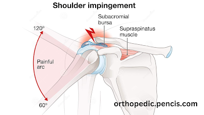.png)
-
 What are joints? Your joints are places where two or more bones come together. Your shoulders, elbows, hips, knees, and knuckles are all joi...
 -
.jpg) If you've sprained your ankle, you know what severe pain is. But maybe that "sprain" was a "strain" or possibly even...
If you've sprained your ankle, you know what severe pain is. But maybe that "sprain" was a "strain" or possibly even... -
%20(8).png) Golfer's elbow is a condition that causes pain where the tendons of your forearm muscles attach to the bony bump on the inside of your e...
Golfer's elbow is a condition that causes pain where the tendons of your forearm muscles attach to the bony bump on the inside of your e...