Monday, March 13, 2023

Artificial intelligence as good as other methods for documenting hand surgery cases


Automatic-populated clinical notes that are artificial intelligence-based may decrease documentation burden for hand surgery cases in the future vs. transcription services or voice recognition systems, according to a presenter.

In research presented at the American Academy of Orthopaedic Surgeons Annual Meeting, Michael Rivlin, MD, FAAOS, discussed results of a prospective study he and his colleagues conducted to compare the quality of documentation of common orthopedic encounters for patients who undergo hand surgery with an artificial intelligence (AI)-based virtual scribe service, a transcription service and a voice recognition mobile (VRM) application.

Provider time is a critical resource that needs to be conserved, particularly with the high rates of physician mental health issue and burnout today, Rivlin told Healio in an interview.

“The more provider time you can give back to the provider, the more they can spend on caring for patients, so modalities, such as AI scribe or an in-person scribe, are helpful, but over time we expect the AI scribe to replace the human factor,” Rivlin said.

Because the quality of AI-generated documentation had not been analyzed previously, Rivlin and colleagues compared the quality and time spent documenting common orthopedic encounters in hand surgery, according to a press release. They used the following modalities during a patient visit:

AI-based virtual scribe service, which is an AI program that runs on a tablet and a machine extracts everything said in the room;

medical scribe, which a person either physically in the office visit or who participates virtually and transcribes the entire patient encounter;

transcription service, in which the physician uses a Dictaphone device to record an audio file about the patient visit and sends it to a third-party company so that what has been dictated can be transcribed; and

VRM application, which is a program available on electronic medical record platforms that types the words said based on voice recognition.


According to a press release for the study, three fellowship-trained orthopedic hand surgeons evaluated 10 standardized patients with pre-written clinical vignettes. Physicians not involved in the study acted out the clinical vignettes, which were documented by the different systems being analyzed, according to the press release.

Clinical documentation was performed during the clinical encounter using the AI-based scribe and medical scribe, and then afterward using a VRM and transcription service.

According to the press release, a total of 118 clinical encounters were documented. This included 30 AI scribe, 30 VRM, 28 transcription service and 30 medical scribe notes. Clinical notes were deemed as either acceptable or unacceptable and assigned a letter grade of A, B, C or F using an eight-point scoring system. In addition, an attorney reviewed all notes for medical legal risk.

Overall, all modalities performed well with similar documentation outputs between the modalities. According to the abstract, there were no significant differences in note quality for each hand surgeon who participated in the study, as well as for the entire cohort.

3rd Edition of Orthopedic | 24-26 April 2023 | London, United Kingdom

Award Nomination - https://x-i.me/ortawr21
Award Registration - https://x-i.me/oaregaj5
Member Nomination - https://x-i.me/ortmem0

#Orthopedics#Orthopedic surgery#Joints#Bone#Arthritis#Fracture#Dislocation#Sprain#Strain#Tendinitis
#Bursitis#Osteoporosis

Intraoperative 3D imaging more effective in confirming the accuracy of pedicle screw placement during spine surgery

A study at Hospital for Special Surgery (HSS) found that intraoperative three-dimensional (3D) imaging was superior to two-dimensional radiographs in confirming the accuracy of pedicle screw placement during spine surgery. The research was presented today at the American Academy of Orthopaedic Surgeons (AAOS) Annual Meeting in Las Vegas.

Many spinal surgeries require the use of implants called pedicle screws to stabilize the spine. Precise positioning of these screws is critical for a successful outcome.

Dr. Lebl and colleagues set out to compare the accuracy of BPR versus 3D imaging when assessing intraoperative pedicle screw placement. "Our study is the first to compare the differences in intraoperative biplanar radiography and 3D imaging for pedicle screw accuracy in thoracic and lumbar cases using robotic technology," Dr. Lebl noted.

Investigators analyzed data from 103 patients who underwent spinal fusion by a single surgeon from 2019 to 2022. Pedicle screw placement was assessed with both intraoperative BPR and 3D imaging in each case.

"CT scans taken after surgery were compared to the findings of intraoperative BPR and 3D imaging to detect either false-positive or false-negative readings," explained Fedan Avrumova, BS, an HSS clinical research coordinator who presented the study at the AAOS meeting. "False positive findings are instances when BPR imaging suggests the screw was not in an acceptable position, while in fact a more advanced 3D image (intraoperative 3D scan or postoperative CT scan) showed the screw to be in an acceptable position. Conversely, a false negative instance was when a BPR image led one to believe or looked as though the screw was in an acceptable position, when in fact a more advanced 3D image or post-operative CT scan showed that it was in fact not acceptable."


Postoperative CT imaging revealed a clinically significant number of patients who had false-negative and false-positive screw placement readings on BPR. However, screw position shown on intraoperative 3D imaging was found to be much more accurate, Avrumova added.

"Based on our study, BPR imaging may lead one to think a screw is acceptable when in fact it is not, and also may miss many screws that are not in fact acceptable. In our study, it was approximately one percent of cases where this occurred. However, for surgeons and centers that implant hundreds and thousands of screws per year, this is going to result in a significant clinical impact for many people," Dr. Lebl noted. "Even one misplaced screw can have a significant impact for a patient, a surgeon, and a hospital system. Therefore, based on these findings, we suggest that for intraoperative confirmation of screw position 3D imaging may soon represent a new standard of care.


International Conference on Orthopedics and Sports Medicine


Speaker: Lidocaine has more favorable complication profile for neuraxial anesthesia in THA


Compared with bupivacaine, lidocaine had a more favorable complication profile when used for neuraxial anesthesia in patients undergoing total hip arthroplasty, according to presented results.

“Many people are moving toward regional anesthesia, such as neuraxial anesthesia (NA) [for THA],” Robert Scott Roundy, BS, said in his presentation at the American Academy of Orthopaedic Surgeons Annual Meeting. “However, what type of anesthetic to use in NA is still being debated. One of the worst complications of NA is transient neurological symptoms (TNS) or postoperative pain in the buttocks or extremities


Roundy and colleagues from Emory University performed a prospective, double-blinded randomized controlled trial that compared outcomes of 54 patients who received bupivacaine and 51 patients who received lidocaine for NA during THA. According to the study abstract, outcomes included TNS, time to normal sensation, urinary retention, ambulation and length of stay.

Overall, Roundy and colleagues found no differences in time to normal sensation, urinary retention, ambulation or length of stay between the groups. They noted patients in 13 patients (24%) in the bupivacaine group had urinary incontinence compared with no patients in the lidocaine group. Additionally, seven patients (13%) in the bupivacaine group reported having difficulty with urination or defecation compared with no patients in the lidocaine group.

“These data support the use of lidocaine as part of NA in total hips.

He noted that with increasing rates of outpatient surgery, as well as decreasing OR times and costs of lidocaine, this treatment may have further benefits that were not captured in this study.

3rd Edition of Orthopedic | 24-26 April 2023 | London, United Kingdom

Award Nomination - https://x-i.me/ortawr21
Award Registration - https://x-i.me/oaregaj5
Member Nomination - https://x-i.me/ortmem0


Arthroplasty surgeons showed more physiological stress, strain in revision vs. primary TJA

Results presented at the American Academy of Orthopaedic Surgeons Annual Meeting showed arthroplasty surgeons experienced significantly greater physiological stress and strain when performing revision total joint arthroplasty.

“When scheduling revision and primary total joint arthroplasty, surgeons should consider the higher physiological demand associated with revision total joint arthroplasty and ensure adequate personal preparation,” Irfan A. Khan, ATC, said in his presentation here. “Further study is needed to determine the generalizability of these results and to identify modifiable risk factors for stress and strain during arthroplasty.”

Khan and colleagues collected cardiorespiratory data using a smart vest among two high-volume fellowship-trained arthroplasty surgeons during primary (n=35) and revision (n=35) TJA.


Researchers collected patient BMI, surgical limb laterality and operative time, along with surgeon heart rate, surgeon stress index and surgeon heart rate variability.


Khan noted revision TJA cases had significantly lower BMI, significantly longer operative time and were more often performed using the posterolateral approach.

“When looking at differences between primary and revision total joint arthroplasty, there was a significantly higher energy expenditure and calories per hour, as well as significantly higher heart rate when performing revision total joint arthroplasty.

He added surgeons experienced a significantly higher rate of stress when performing revision TJA.

In subgroup analysis, revision TKA was associated with a significantly higher energy expenditure, heart rate and stress index, according to Khan. He added revision TKA was linked with a significantly lower heart rate variability, indicating increased stress and strain compared with primary TKA.

“When doing a subgroup analysis for hip cases, there were no significant differences for patient BMI or operative time; however, there was a significantly higher rate of energy expenditure when performing revision total hip arthroplasty and, although it was not significant, there does appear to be a higher stress index as well when performing a revision total hip arthroplasty.

3rd Edition of Orthopedic | 24-26 April 2023 | London, United Kingdom


More information :https://orthopedic-conferences.pencis.com/


#Arthroplasty surgeons#Orthopedics#Orthopedic surgery#Joints#Bone#Arthritis#Fracture#Dislocation#Sprain#Strain#Tendinitis
#Bursitis#Osteoporosis

Sling immobilization may be optional after open Latarjet surgery for shoulder instability

LAS VEGAS — Presented results found similar functional and pain outcomes between patients with and those without sling immobilization after open Latarjet surgery for shoulder instability.

“In [patients who undergo open Latarjet surgery], you want to avoid stiffness, especially in external rotation. You want to avoid muscular atrophy, [and] you want to return them to their daily activities as early as possible,” Patrick Goetti Sr., MD, said in his presentation at the American Academy of Orthopaedic Surgeons Annual Meeting. “But we do not know if there is a predictive effect of immobilization on complication rates,” he said.


Goetti and colleagues performed a randomized controlled trial that analyzed functional and pain outcomes of 72 patients with anterior shoulder instability who underwent an open Latarjet procedure either with or without sling immobilization. Outcome measures included Rowe, single assessment numeric evaluation (SANE) and VAS pain scores at baseline, 1.5-, 3- and 6-month postoperative timepoints. Goetti noted grafts were secured with two, threaded 4-mm cancellous screws, which were 1 cm apart from each other.

At 6 months, both groups had showed significant improvements in all outcomes. Mean Rowe scores improved from 38.8 to 81.6; mean SANE scores improved from 42.5 to 84.7; and mean VAS scores improved from 27.7 to 13.9.

Researchers found no significant differences in outcomes between the groups. Mean Rowe scores were 80.7 in the sling group and 82.6 in the no-sling group; mean SANE scores were 83.7 in the sling group and 85.7 in no-sling group; and mean VAS scores were 15.6 in the sling group and 12.2 in the no-sling group. Goetti also noted CT evaluation revealed no differences in graft healing between the groups.

“The absence of postoperative immobilization did not increase complications rates,” Goetti said. “We therefore modified our practice and stopped using sling immobilization after open Latarjet,” he concluded.

#Orthopedics#Orthopedic surgery#Joints#Bone#Arthritis#Fracture#Dislocation#Sprain#Strain#Tendinitis
#Bursitis#Osteoporosis#Scoliosis#Kyphosis#Lordosis

Saturday, March 11, 2023

Trauma surgery

Trauma surgery is the specialization in surgery that focuses on the treatment and care of injuries, often life-threatening, that are caused by impact forces. The causes of impact forces are many, but some of the more common ones include traffic accidents, falls, sports and crush injuries, as well as gunshot or stabbing wounds.

In catastrophic incidents, trauma surgeons often form part of a larger team of specialized surgeons such as orthopedic (i.e. specialist for musculoskeletal injuries), vascular (i.e. specialist for the arteries, veins and other vessels), maxillofacial (i.e. specialist for facial injuries), cardiothoracic (i.e. specialist for the heart and thoracic organs), plastic (i.e. special for the reconstruction of body areas following) and neurosurgeons (i.e. specialists for injuries to the brain and nervous system).

Trauma surgery is a fast-paced and demanding practice that has very little time for the lengthy discussions that may otherwise be seen in some medical consultations. The trauma surgeon generally undergoes training after completion of a basic medical degree.

This training together with the medical degree may take up to a decade or more in most countries. Moreover, in some jurisdictions, trauma surgeons may also conduct the duties that would otherwise be done by general surgeons. This discipline, when combined with urgent general surgery may be referred to as acute surgery care.Trauma surgery is the specialization in surgery that focuses on the treatment and care of injuries, often life-threatening, that are caused by impact forces. The causes of impact forces are many, but some of the more common ones include traffic accidents, falls, sports and crush injuries, as well as gunshot or stabbing wounds.

In catastrophic incidents, trauma surgeons often form part of a larger team of specialized surgeons such as orthopedic (i.e. specialist for musculoskeletal injuries), vascular (i.e. specialist for the arteries, veins and other vessels), maxillofacial (i.e. specialist for facial injuries), cardiothoracic (i.e. specialist for the heart and thoracic organs), plastic (i.e. special for the reconstruction of body areas following) and neurosurgeons (i.e. specialists for injuries to the brain and nervous system).

Trauma surgery is a fast-paced and demanding practice that has very little time for the lengthy discussions that may otherwise be seen in some medical consultations. The trauma surgeon generally undergoes training after completion of a basic medical degree.


This training together with the medical degree may take up to a decade or more in most countries. Moreover, in some jurisdictions, trauma surgeons may also conduct the duties that would otherwise be done by general surgeons. This discipline, when combined with urgent general surgery may be referred to as acute surgery care.

Procedures involved in trauma surgery

In most jurisdictions, trauma surgeons are adequately trained and equipped to identify and handle injuries to the head and neck, abdominal area, chest, legs, feet, arms and hands. Any patient that has experienced traumatic physical injury may be seen by a trauma specialist.

Upon arrival to the emergency room, patients are quickly assessed to identify the extent of the injuries and which are the most threatening to function and life. Resuscitation and stabilization are key priorities prior to surgical operations if urgent surgery is not necessary to save the patient’s life. This is then followed by definitive surgical therapy.

Methods used to assess the extent of injury include radiographic X-rays and CT-scans as well as MRI’s. With these tools the surgeon is able to identify damage to internal organs and hemorrhaging. Trauma surgeons work closely with emergency staff in the resuscitative and stabilization efforts of the patient.

Airway patency, breathing, circulation and necessary drugs are the key parameters controlled. Triage care at admission checks typical details, such as the patient’s vital signs, age and history or pre-existing conditions, like cardiopulmonary diseases. Laboratory tests, like blood tests, may be necessary, as well as intravenous access lines and equipment for monitoring vital signs.

3rd Edition of Orthopedic | 24-26 April 2023 | London, United Kingdom


More information :https://orthopedic-conferences.pencis.com/

#Orthopedics#Orthopedic surgery#Joints#Bone#Arthritis#Fracture#Dislocation#Sprain#Strain#Tendinitis
#Bursitis#Osteoporosis#Scoliosis#Kyphosis#Lordosis
#Spine#Back pain#Neck pain
#Hip#Knee#Shoulder
#Elbow#Wrist#Hand#Foot#Ankle#Joint replacement

                                                                For Enquiries
                                               orthopedic@pencis.com

Thursday, March 9, 2023

how many bones in face

The facial skeleton consists of 14 bones with different anatomic structures and embryological formations (2 unpaired and 6 paired). These bones comprise the paired nasal bones, inferior nasal conchae, palatine bones, maxillae, zygomatic bones, lacrimal bones, and the unpaired mandible and vomer.


Skull bones – Facial and Cranial Bones

The skull comprises numerous, separate bones fused at the immobile joints, referred to as sutures, except for the mandible, which is fused to the skull by the mobile, synovial Temporomandibular joints (TMJ).

Normally, the human skull has twenty-two bones – fourteen facial skeleton bones and eight cranial bones.

The space containing the brain is the cranial cavity. The calvarium or the skull vault is the upper part of the cranium, forming the roof and the sidewalls of the cranial cavity. The foundation of the skull is the lower part of the cranium, constituting the floor of the cranial cavity.

What are the 22 Bones of the Skull?
The skull bones can be broadly divided into two main sections –

       Facial skeleton bones (14)

       Cranial bones (8)

Human Skeletal System

The skeletal system functions as the basic framework of a body and the entire body are built around the hard framework of Skeleton. It is the combination of all the bones and tissues associated with cartilages and joints. Almost all the rigid or solid parts of the body are the main components of the skeletal system. Joints play an important role in the skeletal system as it helps in permitting the different types of movements at different locations. If the skeleton were without joints, then there would be no sign of the movements in the human body.

3rd Edition of Orthopedic | 24-26 April 2023 | London, United Kingdom

Visit:https://orthopedic-conferences.pencis.com/
#Orthopedics#Orthopedic surgery#Joints#Bone#Arthritis#Fracture#Dislocation#Sprain#Strain#Tendinitis
#Bursitis#Osteoporosis#Scoliosis#Kyphosis#Lordosis
#Spine#Back pain#Neck pain
#Hip#Knee#Shoulder
#Elbow#Wrist#Hand#Foot#Ankle#Joint replacement

                                       
                                                          For Enquiries
                                                   orthopedic@pencis.com


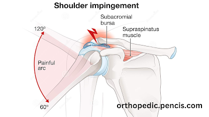
Impingement

Impingement refers to a medical condition characterized by the compression or pinching of soft tissues, such as tendons or bursae, between ...