Saturday, May 13, 2023

Impingement


Impingement refers to a medical condition characterized by the compression or pinching of soft tissues, such as tendons or bursae, between bones in a joint. It commonly occurs in the shoulder, although it can also affect other joints in the body, such as the hip or knee.

In the case of shoulder impingement, the tendons and bursae in the shoulder can become compressed or irritated when there is inadequate space between the upper arm bone (humerus) and the acromion, which is part of the shoulder blade (scapula). This can lead to pain, inflammation, and restricted movement.

Causes of shoulder impingement can vary, but some common factors include:

Overuse or repetitive overhead activities: Activities that involve repetitive overhead motions, such as throwing, swimming, or lifting weights, can increase the risk of impingement.


Structural abnormalities: Certain anatomical variations, such as a curved or hooked acromion, may reduce the space available for the tendons to move freely.


Muscle imbalances or weakness: Weakness or imbalances in the muscles around the shoulder joint can affect the stability and proper alignment of the joint, contributing to impingement

    Symptoms of shoulder impingement may include:

    Pain or tenderness in the shoulder, especially with overhead activities or reaching behind the back.

    Limited range of motion or difficulty with certain movements.

    Weakness or loss of strength in the affected arm.

    Swelling or inflammation around the shoulder joint.


    Treatment options for shoulder impingement depend on the severity of the condition but may include:
    Rest and activity modification: Avoiding activities that aggravate the symptoms and allowing the shoulder to rest and heal.
Physical therapy: Exercises and stretches to improve shoulder strength, flexibility, and posture.

Pain relief measures: Nonsteroidal anti-inflammatory drugs (NSAIDs), ice packs, or corticosteroid injections to reduce pain and inflammation.

Lifestyle modifications: Adjustments in posture, ergonomics, or technique for sports or activities to reduce strain on the shoulder joint.

Surgical intervention: In severe cases that do not respond to conservative treatments, surgery may be considered to create more space in the shoulder joint or repair damaged tissues.

It's important to consult with a healthcare professional, such as a doctor or physical therapist, for an accurate diagnosis and appropriate treatment plan tailored to your specific condition.


Impingement can refer to various medical conditions where tissues in the body become compressed or pinched, leading to pain, inflammation, and restricted movement. While I provided information specifically about shoulder impingement in my previous response, impingement can occur in other areas of the body as well. Here are a few examples:

Shoulder Impingement: This occurs when the tendons or bursae in the shoulder joint are compressed or irritated, typically due to the narrowing of the space between the upper arm bone and the acromion process of the shoulder blade.


Hip Impingement: Also known as femoroacetabular impingement (FAI), this condition involves abnormal contact between the ball-shaped head of the femur (thigh bone) and the socket (acetabulum) in the hip joint. It can result in pain, limited range of motion, and damage to the hip joint cartilage.


Ankle Impingement: Ankle impingement occurs when there is compression or pinching of soft tissues in the ankle joint, often caused by bony growths or excessive scar tissue. It can lead to pain, swelling, and difficulty with walking or movement.


Spinal Impingement: Spinal impingement, also known as nerve impingement or a pinched nerve, occurs when a nerve root in the spinal column is compressed or irritated. This can result in pain, numbness, tingling, or weakness along the path of the affected nerve.

These are just a few examples of impingement conditions, and there may be others depending on the specific area of the body involved. Treatment options for impingement conditions generally focus on relieving pain, reducing inflammation, improving range of motion, and addressing any underlying causes. It's essential to consult with a healthcare professional for an accurate diagnosis and appropriate treatment plan tailored to your specific situation.

visit:https://orthopedic-conferences.pencis.com/

#impingement, #shoulderimpingement, #hipimpingement

Monday, May 8, 2023

Bankart repair with remplissage may lower recurrent dislocation rate in Hill-Sachs lesions

Bankart repair with remplissage is a surgical technique used to address recurrent shoulder dislocations in the presence of Hill-Sachs lesions. A Hill-Sachs lesion is a compression fracture of the humeral head that can occur during a shoulder dislocation.

During a Bankart repair, the torn labrum (a ring of cartilage that surrounds the glenoid) is repaired or reattached to the glenoid using sutures. However, in cases where a large Hill-Sachs lesion is present, there is a risk of the humeral head engaging with the glenoid defect and causing recurrent instability.

To address this issue, the remplissage procedure is performed in conjunction with the Bankart repair. In remplissage, the infraspinatus tendon (a muscle in the back of the shoulder) is transferred and placed into the Hill-Sachs defect, effectively filling the defect and creating a barrier to engagement.

This combined approach of Bankart repair with remplissage has been shown to lower the recurrent dislocation rate in cases with significant Hill-Sachs lesions. The presence of remplissage helps prevent engagement of the humeral head in the glenoid defect, reducing the risk of subsequent dislocations.

Several studies have reported positive outcomes with the Bankart repair and remplissage technique, demonstrating improved stability and reduced rates of recurrent dislocation in patients with significant Hill-Sachs lesions.

However, it's important to note that the decision to perform Bankart repair with remplissage depends on various factors, including the size and severity of the Hill-Sachs lesion, patient characteristics, and surgeon expertise. A thorough evaluation by an orthopedic surgeon is necessary to determine the most appropriate surgical approach for each individual case.

In summary, Bankart repair with remplissage is a surgical technique that combines the repair of a torn labrum with the filling of a Hill-Sachs lesion to enhance stability and reduce the risk of recurrent shoulder dislocations. It can be an effective treatment option for patients with significant Hill-Sachs lesions and recurrent instability.


Bankart repair is a surgical procedure performed to address shoulder instability, specifically anterior shoulder instability. It is named after the surgeon who first described the technique, Dr. Arthur Bankart.

Anterior shoulder instability occurs when the humeral head (upper arm bone) dislocates or partially dislocates from the glenoid (shoulder socket) in a forward direction. This can result in recurrent shoulder dislocations and a feeling of instability.

During a Bankart repair, the torn labrum (a ring of cartilage that surrounds the glenoid) is repaired or reattached to the glenoid using sutures. The labrum acts as a stabilizing structure for the shoulder joint, and repairing it helps restore stability and prevent further dislocations.

The surgery is typically performed arthroscopically, using small incisions and a camera (arthroscope) to guide the procedure. The surgeon inserts specialized instruments through additional small incisions to repair the labrum and address any associated soft tissue damage.

Bankart repair is often accompanied by other procedures, depending on the specific condition of the patient's shoulder. These additional procedures may include addressing any capsular laxity (loose ligaments) or addressing other associated injuries such as Hill-Sachs lesions (compression fractures on the humeral head).

Following the surgery, a period of rehabilitation and physical therapy is usually required to restore strength, range of motion, and stability to the shoulder joint. The length and intensity of the rehabilitation process will vary depending on the individual and the extent of the initial injury.

Bankart repair is considered a successful surgical intervention for anterior shoulder instability, with a high rate of returning patients to their pre-injury level of activity and reducing the risk of recurrent dislocations. However, the success of the surgery can depend on various factors, including the severity of the initial injury, patient compliance with rehabilitation, and the surgeon's skill and experience.

It is important for individuals considering Bankart repair or any surgical procedure to consult with an orthopedic surgeon who can provide a thorough evaluation and determine the most appropriate treatment plan based on their specific condition.


Follow us to learn more about orthopedic surgeries and advancements in shoulder treatments. #BankartRepair #ShoulderStability #Orthopedics"#BankartRepair #ShoulderSurgery #ShoulderStability #Orthopedics #OrthopedicSurgery #SportsInjuries #ShoulderInstability #ShoulderRehabilitation

Nonrigid fixation may yield higher failure rates after arthroscopic glenoid reconstruction

Nonrigid fixation in arthroscopic glenoid reconstruction refers to the use of techniques that do not provide rigid stability or secure fixation of the glenoid bone. This can include methods such as suture anchors or soft tissue grafts.

Several studies and research have suggested that nonrigid fixation in arthroscopic glenoid reconstruction may result in higher failure rates compared to rigid fixation techniques. Failure in this context refers to recurrent instability or inadequate healing of the reconstructed glenoid.

One reason for the higher failure rates with nonrigid fixation is the potential for insufficient initial stability of the reconstructed glenoid. Without rigid fixation, there is a greater likelihood of graft or anchor loosening, leading to recurrent instability or graft failure.

Rigid fixation techniques, on the other hand, provide more secure and stable fixation of the glenoid, allowing for better healing and reduced risk of recurrent instability. These techniques typically involve the use of screws or other hardware to secure the bone graft or anchor in place.

It's important to note that the choice of fixation technique in arthroscopic glenoid reconstruction depends on various factors, including the patient's specific condition, surgeon expertise, and other anatomical considerations. The decision should be made based on a thorough evaluation of the individual case.

Overall, while nonrigid fixation techniques may have some advantages in certain cases, they should be used cautiously, and the potential for higher failure rates should be carefully considered and discussed with the surgeon. Rigid fixation techniques generally remain the gold standard for achieving long-term stability and successful outcomes in arthroscopic glenoid reconstruction.


Nonrigid fixation refers to the use of techniques that do not provide rigid stability in arthroscopic glenoid reconstruction, a surgical procedure used to repair and stabilize the glenoid (shoulder socket). Instead of using rigid hardware like screws, nonrigid fixation methods utilize sutures or soft tissue grafts to secure the glenoid.

Recent research suggests that nonrigid fixation may lead to higher failure rates compared to rigid fixation techniques. Failure in this context refers to recurrent instability or inadequate healing of the reconstructed glenoid.

One of the reasons behind the higher failure rates associated with nonrigid fixation is the potential for inadequate initial stability. The absence of rigid fixation can result in increased movement or loosening of the graft or anchor, leading to instability and compromised healing.

Rigid fixation techniques, such as the use of screws or other hardware, offer more secure and stable fixation of the glenoid. This allows for better healing and reduces the risk of recurrent instability or graft failure.

It's essential to note that the choice between rigid and nonrigid fixation depends on various factors, including the specific case, patient characteristics, and surgeon preference. There may be situations where nonrigid fixation is appropriate, such as in certain anatomical variations or cases where rigid fixation is not feasible.

The decision on which fixation technique to use should be made on a case-by-case basis, considering factors such as the patient's condition, surgeon expertise, and the desired outcome. It is important for patients to have a thorough discussion with their orthopedic surgeon to understand the pros, cons, and potential risks associated with both rigid and nonrigid fixation options.

In summary, while nonrigid fixation techniques have certain advantages, such as versatility and potential for less invasive surgery, they may carry a higher risk of failure compared to rigid fixation methods. Surgeons carefully evaluate each case to determine the most appropriate fixation technique that will provide optimal stability and long-term success for arthroscopic glenoid reconstruction.


#NonrigidFixation #ArthroscopicGlenoidReconstruction #ShoulderSurgery #Orthopedics #OrthopedicResearch #JointReconstruction #SurgicalFixation #ShoulderInjury #OrthopedicFailures #GlenoidInstability

Bone and joint infections


In this educational video, we delve into the world of bone and joint infections. From common conditions like osteomyelitis to septic arthritis, we explore the causes, symptoms, diagnosis, and treatment options for these musculoskeletal infections. Join us as we discuss the latest advancements in orthopedic infectious disease management, highlighting both surgical and non-surgical approaches. Stay informed and gain valuable insights into how healthcare professionals handle bone and joint infections.

  Follow for more orthopedic education and stay updated with the latest advancements in the field.

#bone infection,# joint infection, #orthopedic infections,# osteomyelitis, #septic arthritis, #musculoskeletal #infections, #orthopedics, #infectious diseases, #treatment, #diagnosis

Friday, May 5, 2023

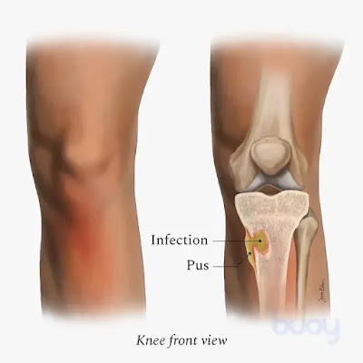
Bone & Joint Infections


If bacteria enter the body, they can find their way to the musculoskeletal system, leading to infection. Bone and joint infections, also known as musculoskeletal infections, occur most commonly in toddlers and young children. They often occur in the bones and joints of the arms and legs. An infection of the bone is called osteomyelitis and septic arthritis refers to a joint infection.
Bone and joint infections are usually caused by bacteria called Staphylococcus aureus (or “staph”) and require treatment with antibiotics. A serious form of staph known as methicillin-resistant Staphylococcus aureus (MRSA) causes infections that can be more severe and need special antibiotics to treat.

It is important to recognize and treat bone and joint infections because they can:


Spread to the blood and other organs causing life-threatening illness
Damage growth plates, bones and joints
Cause chronic arthritis and bone fractures

Symptoms of bone and joint infections may include:

Pain (localized or vague) in a limb or joint that increases in severity
Pain with bearing weight on the affected limb or joint
Pain with touching the affected area
Redness and swelling at the site of pain
Redness and swelling of the joint
Warmth and redness in the infected area
Fever
Fatigue
General malaise

Diagnosis of Bone & Joint Infections

Pediatric infectious disease specialists at Riley at IU Health diagnose children with symptoms of an infection using the following tests:

Physical exam. This is a routine test that doctors use to assess health.
Blood test. This test examines a sample of blood.
X-ray. This test produces images of the inside of the body to help identify possible areas of infection.
Computed tomography (CT). This special X-ray produces cross-sectional images of the body to see if certain areas of the body are infected.
Magnetic resonance imaging (MRI). This test produces images of the inside of the body to see if there is inflammation in the infected areas. Some children may need diagnostic and interventional anesthesia for this test.

Once an experienced pediatrician or pediatric orthopedist determines that your child has a bone or joint infection, a procedure may be required to obtain a sample from the infected area. In the case of joint infections, fluid is aspirated using a needle and sent for laboratory analysis. Infections of the bone may require a bone biopsy (tissue sample taken from the body and examined) to help identify what specific bacteria have infected the bone. These procedures help decide the appropriate antibiotic for that type of bacterial infection. Occasionally, orthopedic surgery is required in cases of complicated musculoskeletal infections.

Treatments

If your child has a bone or joint infection, he or she needs to be treated by an experienced pediatric infectious disease specialist as well as a pediatric orthopedic surgeon. If infections are identified and treated early, most children have a good outcome.

Bone and joint infections are treated with intravenous (IV) antibiotics during a short hospital stay. After leaving the hospital, most children take oral antibiotics, usually for several weeks.

Some children may need a special IV called a peripherally inserted central catheter (PICC) line. A PICC line provides intravenous access for a prolonged period of time so the child can continue to receive IV antibiotics at home.

Treatment should include follow-up with a pediatric infectious disease specialist and a pediatric orthopedist.


Bone and joint infections, also known as musculoskeletal infections, occur most commonly in toddlers and young children.
Bone and joint infections are usually caused by bacteria called Staphylococcus aureus (or “staph”) and require treatment with antibiotics.
When treated early, most children with bone and joint infections have a positive outcome.
Some complicated cases of musculoskeletal infections may require orthopedic surgery.

Support Services & Resources

Find more information about award-nomination by visiting these trusted websites.


Nomination Link: https://x-i.me/ortawr21

Bone & Joint Infections Research

The Infectious Diseases Department and the Radiology & Imaging Department at Riley at IU Health are conducting research on the most effective diagnostic methods for musculoskeletal infections.


visit:https://orthopedic-conferences.pencis.com/

#BoneInfections #JointInfections #Osteomyelitis #SepticArthritis #Discitis #Bursitis #InfectionPrevention #Antibiotics #Surgery #Microorganisms #MedicalConditions #Diagnosis #Treatment #RiskFactors #WoundCare #Hygiene #IntravenousDrugUse #WeakenedImmuneSystem #MRI #Xray #CTscan #BloodTests #Pus #Abscesses #InfectedTissue #Complications #LongTermDisability

Surgery for scoliosis

Scoliosis is a condition that causes the spine to curve to the side, resulting in an abnormal posture and sometimes causing discomfort or difficulty breathing. While some cases of scoliosis can be managed through non-surgical means such as bracing or physical therapy, others may require surgery to correct the curvature of the spine.

Surgery for scoliosis typically involves a procedure called spinal fusion, which aims to fuse the affected vertebrae together so that they can no longer move independently. During the surgery, the surgeon will use metal rods, screws, and other devices to straighten the spine and hold it in place while the bones fuse together over time.

Spinal fusion surgery for scoliosis is a complex procedure that carries some risks, including infection, bleeding, and nerve damage. However, for many people with severe scoliosis, surgery can be an effective way to improve their quality of life and prevent further progression of the condition.

Recovery from scoliosis surgery can be lengthy and may involve a period of immobilization or limited activity. Physical therapy and rehabilitation are often recommended to help restore mobility and strength in the affected area. Your doctor will work with you to develop a personalized treatment plan based on your individual needs and the severity of your scoliosis.


Surgery for scoliosis is usually recommended when the curvature of the spine is severe (typically greater than 40-50 degrees), causing significant pain or discomfort, or interfering with daily activities such as breathing or walking.


The goal of scoliosis surgery is to correct the curvature of the spine and prevent further progression of the condition. This is typically achieved through spinal fusion, which involves fusing together the affected vertebrae using metal rods, screws, or other devices.


Recovery from scoliosis surgery can be lengthy and may involve a period of immobilization or limited activity. Physical therapy and rehabilitation are often recommended to help restore mobility and strength in the affected area.


Types of scoliosis surgery:

Posterior spinal fusion: This is the most common type of scoliosis surgery, in which the surgeon makes an incision in the back and uses metal rods, screws, and other devices to straighten the spine and hold it in place while the bones fuse together.


Anterior spinal fusion: In this type of surgery, the surgeon makes an incision in the front of the body and removes a rib to access the spine. Metal devices are used to straighten the spine and hold it in place while the bones fuse together.


Thoracoscopic surgery: This is a minimally invasive type of surgery in which the surgeon makes several small incisions in the back and uses a tiny camera and specialized instruments to perform the spinal fusion.


Vertebral body tethering: This is a newer type of surgery that involves attaching a flexible cord to the affected vertebrae to correct the curvature of the spine. Unlike spinal fusion, this procedure allows for some movement in the spine.

It's important to note that the type of surgery recommended for scoliosis will depend on the individual's age, the severity and location of the curvature, and other factors. Your doctor will work with you to determine the best course of treatment based on your individual needs.

Scoliosis surgery is a procedure that can help correct the curvature of the spine in people with severe cases of scoliosis. Spinal fusion is the most common type of surgery for scoliosis, which involves fusing together the affected vertebrae using metal rods, screws, or other devices. Recovery from scoliosis surgery can be lengthy and may involve a period of immobilization or limited activity. Physical therapy and rehabilitation are often recommended to help restore mobility and strength in the affected area.

visit:https://orthopedic-conferences.pencis.com/

 #ScoliosisSurgery #SpinalFusion #RecoveryJourney

What is fracture.


visit https://orthopedic-conferences.pencis.com/


A fracture is a medical condition where a bone is broken. It can be caused by accidents, falls, or repetitive stress on the bone. Symptoms include pain, swelling, and difficulty moving the affected area. Treatment options depend on the severity of the fracture, and may include immobilization with a cast or brace, surgery, pain management, and physical therapy.


#FractureAwareness #BoneHealth #InjuryPrevention

Impingement

Impingement refers to a medical condition characterized by the compression or pinching of soft tissues, such as tendons or bursae, between ...