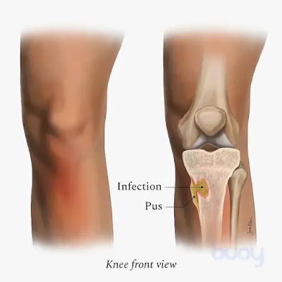
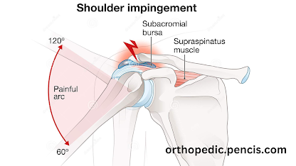
Impingement refers to a medical condition characterized by the compression or pinching of soft tissues, such as tendons or bursae, between bones in a joint. It commonly occurs in the shoulder, although it can also affect other joints in the body, such as the hip or knee.
In the case of shoulder impingement, the tendons and bursae in the shoulder can become compressed or irritated when there is inadequate space between the upper arm bone (humerus) and the acromion, which is part of the shoulder blade (scapula). This can lead to pain, inflammation, and restricted movement.
Causes of shoulder impingement can vary, but some common factors include:
Overuse or repetitive overhead activities: Activities that involve repetitive overhead motions, such as throwing, swimming, or lifting weights, can increase the risk of impingement.
Structural abnormalities: Certain anatomical variations, such as a curved or hooked acromion, may reduce the space available for the tendons to move freely.
Muscle imbalances or weakness: Weakness or imbalances in the muscles around the shoulder joint can affect the stability and proper alignment of the joint, contributing to impingement
Symptoms of shoulder impingement may include:
Pain or tenderness in the shoulder, especially with overhead activities or reaching behind the back.
Limited range of motion or difficulty with certain movements.
Weakness or loss of strength in the affected arm.
Swelling or inflammation around the shoulder joint.
.png)
Treatment options for shoulder impingement depend on the severity of the condition but may include:
Rest and activity modification: Avoiding activities that aggravate the symptoms and allowing the shoulder to rest and heal.
Pain relief measures: Nonsteroidal anti-inflammatory drugs (NSAIDs), ice packs, or corticosteroid injections to reduce pain and inflammation.
Lifestyle modifications: Adjustments in posture, ergonomics, or technique for sports or activities to reduce strain on the shoulder joint.
Surgical intervention: In severe cases that do not respond to conservative treatments, surgery may be considered to create more space in the shoulder joint or repair damaged tissues.
It's important to consult with a healthcare professional, such as a doctor or physical therapist, for an accurate diagnosis and appropriate treatment plan tailored to your specific condition.
Impingement can refer to various medical conditions where tissues in the body become compressed or pinched, leading to pain, inflammation, and restricted movement. While I provided information specifically about shoulder impingement in my previous response, impingement can occur in other areas of the body as well. Here are a few examples:
Shoulder Impingement: This occurs when the tendons or bursae in the shoulder joint are compressed or irritated, typically due to the narrowing of the space between the upper arm bone and the acromion process of the shoulder blade.
Hip Impingement: Also known as femoroacetabular impingement (FAI), this condition involves abnormal contact between the ball-shaped head of the femur (thigh bone) and the socket (acetabulum) in the hip joint. It can result in pain, limited range of motion, and damage to the hip joint cartilage.
Ankle Impingement: Ankle impingement occurs when there is compression or pinching of soft tissues in the ankle joint, often caused by bony growths or excessive scar tissue. It can lead to pain, swelling, and difficulty with walking or movement.
Spinal Impingement: Spinal impingement, also known as nerve impingement or a pinched nerve, occurs when a nerve root in the spinal column is compressed or irritated. This can result in pain, numbness, tingling, or weakness along the path of the affected nerve.
These are just a few examples of impingement conditions, and there may be others depending on the specific area of the body involved. Treatment options for impingement conditions generally focus on relieving pain, reducing inflammation, improving range of motion, and addressing any underlying causes. It's essential to consult with a healthcare professional for an accurate diagnosis and appropriate treatment plan tailored to your specific situation.


%20(1).png)

.png)

.jpg)
%20(8).png)